Article Text

Abstract
BACKGROUND In patients with Peutz-Jeghers syndrome (PJS), causative germline mutations in theLKB1/STK11 gene on chromosome 19p13.3 have been identified. Because of the loss of heterozygosity (LOH) at 19p13.3 in hamartomas and the cancer susceptibility of patients with PJS, LKB1/STK11 is suggested to act as a tumour suppressor. However, the frequency of genetic and epigenetic inactivation ofLKB1/STK11 in sporadic tumours is unclear.
AIMS To investigate the LKB1/STK11 gene for promoter hypermethylation and allelic loss in tumour specimens of patients with sporadic colorectal cancer.
METHODS DNA from 50 consecutive paraffin embedded sporadic colorectal adenocarcinomas and corresponding normal epithelium was extracted. After bisulphite treatment, specimens were analysed for methylation of theLKB1/STK11 promoter 5′-CpG island by methylation specific polymerase chain reaction (MSP). In addition, tumours were analysed for LOH of chromosome 19p13.3. In tumours exhibiting LOH, LKB1/STK11 was sequenced.
RESULTS MSP was successful in 48 of 50 tumour specimens. Of those, four (8%) demonstrated hypermethylation of theLKB1/STK11 promoter 5′-CpG island. Moreover, LOH at either D19S886 or D19S878 was observed in five of 38 (13%) informative tumours. All five tumours showing LOH at 19p13.3 were advanced and four of five were located in the left sided colon. There was no correlation between LOH andLKB1/STK11 promoter hypermethylation or somatic mutation.
CONCLUSIONS In sporadic colorectal cancer, hypermethylation of theLKB1/STK11 promoter and allelic loss at theSTK 11 gene locus are rare events. LOH at 19p13.3 was associated with advanced tumour stage and left sided location but not with LKB1/STK11 promoter hypermethylation or somatic mutation.
- sporadic colorectal adenocarcinoma
- tumour suppressor genes
- protein-serine-threonine kinase geneLKB1/STK11
- promoter hypermethylation
- loss of heterozygosity
Abbreviations used in this paper
- LKB1/STK11
- serine-threonine kinase gene LKB1/STK11
- LOH
- loss of heterozygosity
- PJS
- Peutz-Jeghers syndrome
- UICC
- International Union Against Cancer
- PCR
- polymerase chain reaction
- MSP
- methylation specific polymerase chain reaction
- bp
- base pairs
Statistics from Altmetric.com
- sporadic colorectal adenocarcinoma
- tumour suppressor genes
- protein-serine-threonine kinase geneLKB1/STK11
- promoter hypermethylation
- loss of heterozygosity
Several tumour suppressor genes, which are inactivated through germline mutations in the most commonly inherited colorectal cancer susceptibility syndromes, such as the APCgene in familial adenomatous polyposis and the DNA mismatch repair genes MSH2, MLH1,PMS1, PMS2, andMSH6/GTBP in hereditary non-polyposis colorectal cancer, are involved in the development and progression of sporadic colorectal cancer.1 ,2 Moreover, loss of transcription of tumour suppressor genes, includingp16, MGMT, andMLH1, by epigenetic changes such as hypermethylation of 5′-CpG islands in the promoter region have been demonstrated in colorectal cancer.3 ,4
Recently, a gene mutation in patients with Peutz-Jeghers syndrome (PJS), an autosomal dominant disorder characterised by mucocutaneous pigmentation, intestinal hamartomas, and an increased risk of cancers of the gastrointestinal tract, breast, testis, and ovary, has been identified by genetic linkage studies and positional cloning.5 ,6 This gene, namedLKB1, STK11, orLKB1/STK11, is located on chromosome 19p13.37 and encodes for a serine-threonine kinase, a human homologue of Xenopus early embryonic kinase 1.8 LKB1/STK11 is suggested to act as a tumour suppressor gene in PJS because hamartoma formation in PJS patients with inactivating LKB1/STK11 germline mutations is associated with somatic loss of the wild-typeLKB1/STK11 allele.7 ,9 ,10 The development of cancer in patients with PJS does not exclusively arise in association with hamartomas11 but dysplasia with consecutive neoplastic transformation within hamartomatous polyps accounts for at least some malignancies in this syndrome.12 ,13 In contrast with the pathogenesis of sporadic colorectal cancer, frequently involvingAPC, K-ras,DCC, MCC, andp53,1 ,2 ,14 the molecular mechanisms leading to cancer in patients with PJS remain unclear. Because patients with PJS are at increased risk of colorectal cancer,LKB1/STK11 may also be a target during the carcinogenesis of sporadic colorectal cancer. Although some reports revealed a low frequency of somatic mutations of theLKB1/STK11 gene in colorectal tumour specimens,15-17 conflicting data were most recently reported by Dong and colleagues.18 This group identified somatic LKB1/STK11 mutations in one third of left sided colorectal cancers and in two colonic adenomas. In contrast,LKB1/STK11 promoter hypermethylation leading to transcriptional inactivation was found in a few cancer cell lines and a subset of primary tumours.19
To further analyse the role of LKB1/STK11 on chromosome 19p13.3 in the pathogenesis of sporadic colorectal cancer, we investigated the frequency of genetic and epigenetic alterations at the LKB1/STK11 gene locus in tumour specimens from 50 consecutive patients with colorectal cancer.
Methods
TUMOUR SPECIMENS
Tumour specimens from 50 consecutive patients (21 females, 29 males) with sporadic colorectal adenocarcinoma (International Union Against Cancer (UICC) stage I, n=9; stage II, n=10; stage III, n=17; stage IV, n=14) were analysed. At the time of diagnosis, patients were aged 35–95 years (mean 55 (17) years). In 38 patients tumours were left sided (descending colon, sigmoid colon, or rectum) whereas 12 patients had a right sided colon carcinoma (transverse colon, ascending colon, or caecum). Histologically, two tumours were characterised as well differentiated, 39 as moderately differentiated, six as poorly differentiated, and three as undifferentiated. None of the patients had a family history of PJS, familial adenomatous polyposis, or hereditary non-polyposis colorectal cancer and none of the tumours exhibited microsatellite instability (data not shown).
For molecular analysis, representative 5 μm sections of paraffin embedded normal and tumour tissue were mounted onto slides and dried for 60 minutes at 50°C. After microdissection DNA was extracted using the QIAamp DNA mini kit (Qiagen, Hilden, Germany).
METHYLATION SPECIFIC PCR OF THELKB1/STK11PROMOTER
Analysis of methylation patterns within the 5′-CpG island of the LKB1/STK11 gene were carried out using chemical modification of 1 μg of genomic DNA from colorectal cancer specimens with sodium bisulphite and methylation specific polymerase chain reaction (MSP) using sense primers for methylated and unmethylated polymerase chain reactions (PCR) beginning at base pairs (bp) 15 and 17, respectively, from GeneBank sequence AF 035625, as previously described.19Primers used for the unmethylated reaction were 5′-GGATGA AGTTGATTTTGATTGGGTT-3′ (sense) and 5′-ACCCAATACAAAATCTACAAA CC AACA-3′ (antisense) and for the methylated reaction 5′-ACGAAGTTGATTTTGA TCGGGTC-3′ (sense) and 5′-CGATAC AAAATCTACGAACCGACG-3′ (antisense). PCR was carried out in a final volume of 50 μl containing 3.5 mM magnesium chloride, 15 mM ammonium sulphate, 60 mM Tris HCl (pH 8.5), 250 μM each of dATP, dTTP, dCTP, and dGTP (Invitrogen, Leek, Netherlands), 0.1 μM forward and reverse primers (Biospring, Frankfurt, Germany), and 2.5 U of AmpliTaqGold DNA polymerase (Perkin Elmer, Weiterstadt, Germany) for 10 minutes at 95°C followed by 55 cycles of 30 seconds at 95°C, 30 seconds at 55°C, one minute at 72°C, and a final extension of 10 minutes at 72°C. PCR products were electrophoresed on non-denaturating polyacrylamide gels (8%) and visualised by silver staining.
LOSS OF HETEROZYGOSITY ANALYSIS OF 19p13.3
After PCR amplification of DNA extracted from normal and tumour tissue the microsatellite markers D19S886, located telomeric to theLKB1/STK11 gene locus on chromosome 19p13.3, and D19S878, located 6.5 cM proximal to D19S886,20 were analysed for loss of heterozygosity (LOH). Primer sequences for D19S886 were 5′-TGGATCTACACTCC GGC-3′ (sense) and 5′-ATTTTACTGGC TGGCACTTG-3′ (antisense), and for D19S878 were 5′-GCCTGGGCGACA GAGAAT-3′ (sense) and 5′-GGTTGC CCGCAGAGTG-3′ (antisense). PCR was carried out in a final volume of 50 μl containing 2.5 mM magnesium chloride, 15 mM ammonium sulphate, 60 mM Tris HCl (pH 8.5), 250 μM each of dATP, dTTP, dCTP, and dGTP (Invitrogen), 0.1 μM of 6-carboxy-fluorescein labelled forward and 0.1 μM reverse primers (Biospring), and 2.5 U of AmpliTaq Gold DNA polymerase (Perkin Elmer) for 10 minutes at 95°C followed by 45 cycles of 30 seconds at 95°C, 30 seconds at 55°C, one minute at 72°C, and a final extension of 10 minutes at 72°C. Electrophoresis was carried out in an ABI 310 DNA sequencer (Perkin Elmer) and the final analysis was performed using the Gene Scan 2.1 software (Perkin Elmer).
GENOMIC PCR AMPLIFICATION AND SEQUENCE ANALYSIS OF THELKB1/STK11GENE
Genomic PCR amplification of the coding region of theLKB1/STK11 gene was carried out using published primer sets.5 ,6 ,15 PCR reactions were carried out in a total volume of 50 μl, consisting of 1.5–3.5 mM magnesium chloride, 15 mmol/l ammonium sulphate, 60 mM Tris HCl (pH 8.5), 250 μM each of dATP, dTTP, dCTP, and dGTP (Invitrogen), 1 μM of forward and reverse primers (Biospring), and 2.5 U of AmpliTaq Gold DNA polymerase (Perkin Elmer). The following amplification conditions were used: 12 minutes at 95°C; 40 cycles of 45 seconds at 95°C, one minute annealing at 55°C, and two minutes at 72°C; and a final extension of 10 minutes at 72°C. PCR products of the LKB1/STK11 gene were purified and bidirectionally sequenced according to the instructions of the Dye Deoxy Terminator protocol (Perkin Elmer). Automated sequence analysis was carried out on an ABI 310 DNA sequencer (Perkin Elmer).
Results
LKB1/STK115′-CpG ISLAND METHYLATION
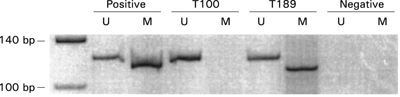
Methylation specific PCR (MSP) for analysis of theLKB1/STK11 promoter 5′-CpG island was carried out with DNA extracted from 50 paraffin embedded colorectal cancer specimens. DNA extracted from the colorectal cancer cell lines HT29 and H6 served as positive controls for the unmethylated and methylated reactions, respectively (fig 1). In two specimens there was insufficient DNA for MSP. In the remaining 48 tumour specimens MSP was successful. Of those, four (8%) tumours demonstrated both methylated and unmethylated LKB1/STK11 promoter 5′-CpG islands whereas the remaining 44 specimens displayed only unmethylated promoter islands. The presence of methylatedLKB1/STK11 promoter 5′-CpG islands was not associated with tumour stage, location, or histological grading.

Methylation specific polymerase chain reaction (PCR) of the LKB1/STK11 promoter 5′-CpG island in sporadic colorectal cancer. In the left lane the 140 bp and 100 bp bands of a molecular weight standard are shown. The presence of a visible PCR product in those lanes marked U indicates unmethylated LKB1/STK11 promoter 5′-CpG islands; a visible PCR product in those lanes marked M indicates the presence of methylated LKB1/STK11 promoter 5′-CpG islands. Corresponding lanes are: positive controls for PCR reactions (for the unmethylated reaction DNA extracted from the colorectal cancer cell line HT29 and for the methylated reaction from the colorectal cancer cell line H6), primary colorectal cancers (T100 and T189), and negative controls for PCR reactions.
LOH ANALYSIS OF 19p13.3
To investigate the LKB1/STK11gene locus in sporadic colorectal cancer for allelic loss at chromosome 19p13.3, we analysed paraffin embedded normal DNA and corresponding tumour DNA. Thirty eight of 50 tumour specimens were considered informative for at least one microsatellite marker. LOH at either D19S886 or D19S878 was observed in five of 38 (13%) informative sporadic colorectal cancer specimens. Allelic loss at D19S878 occurred in four tumours (fig 2) whereas one tumour exhibited LOH at D19S886. Three of five tumours with LOH at 19p13.3 were classified as UICC stage IV whereas the other two were UICC stage III tumours. Four of the five tumours with LOH at 19p13.3 were located in the sigmoid colon or rectum. There was no association between LOH and histological grade. None of the tumours displayingLKB1/STK11 promoter 5′-CpG island hypermethylation was found to exhibit LOH at chromosome 19p13.3.

Fluorescent analysis of the microsatellite marker D19S878 (centromeric to the LKB1/STK11 gene locus) on chromosome 19p13.3. The 123 bp peak of the size standard is plotted in light grey in all electrophoretic profiles. The polymerase chain reaction (PCR) products of normal colon tissue (upper panels) and corresponding tumour tissue (lower panels) in patient No 31 (A) and No 3 (B) with sporadic colorectal adenocarcinoma were electrophoretically analysed on an automated ABI 310 DNA sequencer (Perkin Elmer). (A) Tumour without allelic loss at D19S878. (B) Tumour exhibiting loss of the 127 bp allele of D19S878 whereas the 134 bp allele is conserved.
LKB1/STK11SEQUENCE ANALYSIS
To test if the LKB1/STK11 gene in five tumours exhibiting LOH at 19p13.3 was inactivated because of a somatic mutation of the remaining allele, genomic sequencing of the coding region and splicing sites of the completeLKB1/STK11 gene was performed. In none of those tumours was a somatic LKB1/STK11mutation detected, suggesting that LOH occurred independently of mutational events of the remainingLKB1/STK11 allele.
Discussion
Inactivating LKB1/STK11 germline mutations in combination with loss of the wild-type allele are responsible for the development of hamartomatous polyps in patients with Peutz-Jeghers syndrome (PJS).7 ,9 ,10 To address the pathogenesis of intestinal hamartomas and adenocarcinomas, Gruberet al studied specimens of PJS patients carrying a heterozygote LKB1/STK11 germline mutation.9 Their data further supported the hypothesis that hamartomas and adenocarcinomas in patients with PJS develop through allelic loss of the wild-typeLKB1/STK11 allele whereas alterations of genes affected in the majority of sporadic colorectal carcinomas (APC,K-ras, p53) occur at later stages of tumour progression. ThusLKB1/STK11 is considered a tumour suppressor gene in PJS.
Recently, promoter hypermethylation was established as a new concept of tumour suppressor gene inactivation.3 ,21 In particular, hypermethylation of 5′-CpG islands, usually located in the promoter region of widely expressed genes, has been shown to correlate with decreased or loss of expression. In this study using the published primer sets for the 5′-CpG island of theLKB1/STK11 promoter, we showed the feasibility of LKB1/STK11 promoter methylation analysis in a large series of paraffin embedded colorectal cancer specimens, and confirmed the data of Estelleret al who found one of 43 primary colorectal cancer specimens methylated at theLKB1/STK11 promoter.19
Furthermore, allelic loss at chromosome 19p13.3 was analysed. Using microsatellite markers flanking theLKB1/STK11 gene locus20 we found LOH on chromosome 19p13.3 in five of 38 (13%) informative tumour specimens of patients with sporadic colorectal cancer. To investigate if allelic loss at 19p13.3 occurred in combination with alterations of the remaining allele—according to a “two-hit” inactivation mechanism characteristic of tumour suppressor genes22—we sequenced the entire coding region of theLKB1/STK11 gene in all five tumours with LOH at 19p13.3. No somatic LKB1/STK11 gene mutations were found in these tumours. Our data are in accordance with the studies of Aviziente et al and Restaet al who also did not detect somaticLKB1/STK11 gene mutations in 13 and 10 sporadic colorectal adenocarcinomas with LOH at 19p13.3, respectively.15 ,17 The data are further confirmed by Wanget al who could not detect any bandshifts in the nine exons of the LKB1/STK11 gene in 72 sporadic colorectal adenocarcinomas by single strand conformational polymorphism.16 In contrast, Dong et al detected somatic LKB1/STK11 gene mutations in 7/23 (30%) Korean patients with invasive adenocarcinomas of the sigmoid colon or rectum. Allelic loss at chromosome 19p13.3 was present in six of seven cancers with a somaticLKB1/STK11 mutation.18Moreover, this study reported LKB1/STK11mutations in two of 12 (17%) colonic adenomas with high grade dysplasia. Eight of nine mutations reported by Donget al were missense whereas only one was a frameshift mutation causing a premature stop codon. It remains unclear if these geographical differences inLKB1/STK11 mutation frequency during colorectal tumorigenesis are caused by environmental factors such as dietary exposure to certain carcinogens.
To date, absence or a low frequency of somaticLKB1/STK11 mutations has been reported in sporadic breast cancer (0/62),23 testicular tumour (1/28),15 malignant melanoma (4/85),24-26biliary (1/16), periampullary (0/19),27 and pancreatic adenocarcinoma (5/112),24 ,27 carcinoma of the stomach (3/36),24 ,28 carcinoma of the uterine cervix (1/26), ovarian adenocarcinoma (0/37),29 ovarian granulosa cell tumour (1/24),24 ,29 renal cell cancer (0/19), lung cancer (1/28), and sarcoma (0/24).24 In contrast with the predominance of truncating frameshift germlineLKB1/STK11 mutations in patients with PJS, the vast majority of somatic LKB1/STK11mutations detected so far are missense mutations. Recently, Mehenniet al and Ylikorkala et al reported functional in vitro assays to assess the effects ofLKB1/STK11 germline mutations detected in patients with PJS.30 ,31 Mutant LKB1/STK11 proteins resulting from frameshift or missenseLKB1/STK11 mutations, including a single amino acid change (G163D) reported in a sporadic testicular tumour, resulted in severely impaired or complete loss of kinase activity. Nevertheless, the consequences of somatic missenseLKB1/STK11 mutations have not yet been studied in vivo.
In summary, promoter hypermethylation and allelic loss of theLKB1/STK 11 gene are rare events in sporadic colorectal cancer in Caucasian patients. LOH at 19p13.3 is associated with advanced tumour stage and left sided location but not withLKB1/STK11 promoter hypermethylation or somatic mutation. Because epigenetic or genetic inactivation ofLKB1/STK11 in sporadic colorectal cancer is a rare event, it is unlikely that LKB1/STK11alterations are key players in the molecular pathogenesis of this tumour entity.
Acknowledgments
The authors thank Dr J G Herman, Johns Hopkins Oncology Center, Baltimore, Maryland, USA, for critical review of the manuscript, Dr G Herrmann, Senckenbergisches Zentrum der Pathologie, Klinikum der Johann Wolfgang Goethe-Universität, Frankfurt, Germany, for microdissection of colorectal cancer specimens, and N Weber for technical assistance. This work was supported in part by a grant from the Arthur and Margarete Ebert foundation, Frankfurt, Germany.
Abbreviations used in this paper
- LKB1/STK11
- serine-threonine kinase gene LKB1/STK11
- LOH
- loss of heterozygosity
- PJS
- Peutz-Jeghers syndrome
- UICC
- International Union Against Cancer
- PCR
- polymerase chain reaction
- MSP
- methylation specific polymerase chain reaction
- bp
- base pairs