


Abstract
Background/Aim: We have previously reported that caffeine (CAF) can enhance chemotherapy efficacy of bone and soft-tissue sarcoma established cell lines via cell-cycle perturbation. We subsequently tested the combination of valproic acid (VPA), a histone deacetylase (HDAC) inhibitor, with caffeine on established human osteosarcoma cells in vitro. Both VPA and CAF caused concentration-dependent cell death of the osteosarcoma cell lines in vitro, and their combination was synergistic. We subsequently established patient-derived cell lines from undifferentiated pleomorphic sarcoma (UPS) and rhabdomyosarcoma (RMS), both of which are recalcitrant cancers. These cell lines are termed AC-UPS01 and AC-RMS01, respectively. Materials and Methods: In the present study, we tested CAF and VPA and their combination on the two patient-derived sarcoma cell lines. Cell survival after a 72 h exposure to each drug was determined by the WST-8 assay. IC50 values were calculated for each drug. Results: CAF and VPA caused concentration-dependent cytocidal efficacy for both cell lines. The IC50 for CAF for AC-UPS01 was 2.02 ± 0.22 mM. The IC50 for VPA for AC-UPS01 was 9.54 ± 1.44 mM. The IC50 for CAF for AC-RMS01 was 2.37 ± 0.48 mM. The IC50 for VPA for AC-RMS01 was 2.13 ± 0.20 mM. Synergistic efficacy of combination treatment of CAF and VPA was also observed for both cell lines. Conclusion: The results of the present study suggest that CAF and VPA may be useful in the treatment of recalcitrant sarcoma.
- Undifferentiated pleomorphic sarcoma
- UPS
- rhabdomyosarcoma
- RMS
- patient-derived cell lines
- caffeine
- valproic acid
- cell kill
- synergy
Caffeine (1,3,7-trimethylxanthine) (CAF) has been used to enhance cancer chemotherapy. CAF has several anti-tumor effects, such as apoptosis induction (1). CAF can also modulate the cell cycle of cancer cells. Time-lapse imaging of HeLa cells expressing a fluorescent ubiquitination-based cell cycle indicator (FUCCI) in the nucleus, showed that caffeine increased efficacy of cisplatinum (CDDP) on 143B human osteosaroma cells by increasing the mitotic rate which subsequently increased apoptosis. Caffeine overcame the cell-cycle arrest at S/G2 induced by CDDP, allowing mitosis to proceed, thereby increasing its efficacy (2).
Valproic acid (VPA) is a histone deacetylase (HDAC) inhibitor that also has potential anti-cancer efficacy, shown in clinical trials for leukemia (3), lung cancer (4), and glioblastoma (5, 6).
Sarcomas are a very heterogenetic group of recalcitrant cancers. Previously, we demonstrated cell killing by CAF and VPA and their combination on long-established, high-passage human osteosarcoma cell lines MG63, 143B, and SaOS2 (7). Both VPA and CAF caused concentration-dependent cell death of the osteosarcoma cell lines in vitro. We have also previously reported that CAF-modulation chemotherapy improved the treatment of bone and soft tissue sarcoma in the clinic (8-13).
Previously a patient with high grade undifferentiated pleomorphic soft-tissue sarcoma (UPS) from a striated muscle was implanted orthotopically in the right biceps femoris muscle of mice to establish a patient-derived orthotopic xenograft (PDOX) model (14). A patient-derived orthotopic xenograft (PDOX) mouse model of rhabdomyosarcoma (RMS) was also previously established orthotopically in the right biceps femoris muscle and right quadriceps muscle of nude mice (15).
In the present study, we determined if there was synergistic efficacy of the combination of CAF and VPA against patient-derived UPS and RMS cell lines obtained from the above-described PDOX models.
Materials and Methods
Chemicals. VPA and CAF were obtained from Wako Pure Chemical Industries, Ltd. (Osaka, Japan). CAF and VPA were both dissolved in and diluted with phosphate-buffered saline (PBS). The stock solution for CAF was 25 mM. The stock solution for VPA was 30 mM.
Cell lines. Patient-derived AC-UPS01 and AC-RMS01 cell lines were established from UPS (14) and RMS (15) PDOX mouse models, respectively. The cell lines were grown in RPMI 1640 medium with 10% fetal calf serum. The cell lines were used in passages 5 to 10. Previously, informed-consent was obtained by the patient donors under a UCLA Institutional Review Board-approved protocol (IRB #10-001857) (14, 15).
Growth inhibition assay. Cellular viability was assessed using the WST-8 dye reduction assay. Cells were seeded in 96-well flat-bottomed microplates (100 μl/well) at a 5×10⁴ cells/ml density, incubated at 37°C for 24 h, and exposed to various concentrations of test compounds for 72 h. For each concentration, at least 8 wells were used. After incubation with the test compounds, 10 μl WST-8 solution was added to each well. The microplates were further incubated for 3 h at 37°C, and absorption was measured using a microprocessor-controlled microplate reader (Sunrise™; TECAN, San Joes, CA, USA) at 450 nm. Cell-survival was calculated as the percentage of live cells with respect to untreated control cells. IC50 values were derived from concentration-response curves.
Calculation of Combination Index (CI). The specific interaction between caffeine and valproic acid on the patient-derived UPS and RMS cell lines was evaluated by the combination index (CI) assay using the CalcuSyn software from ComboSyn Inc. (New Jersey, USA) with the method used by Chou and Talalay (16). In this analysis, synergy was defined as a CI <1.0, antagonism as a CI >1.0, and additivity as CI values not significantly different from 1.0.
Results and Discussion
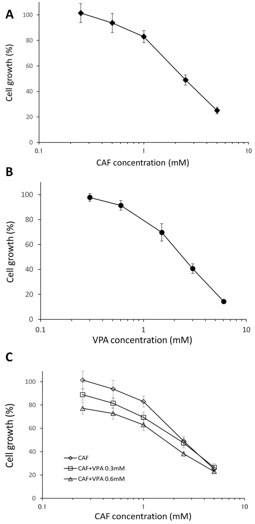
Efficacy of caffeine (CAF) and valproic acid (VPA) on patient-derived undifferentiated pleomorphic sarcoma (UPS) and rhabdomyosarcoma (RMS) cell lines. The cytotoxic activity of CAF and VPA was determined on the patient-derived UPS (AC-UPS01) and RMS (AC-RMS01) cell lines. Cells were incubated for 72 h with each compound. Cell survival was evaluated as described in the Materials and Methods. CAF and VPA significantly inhibited the patient-derived UPS (Figure 1) and RMS (Figure 2) cell lines in a dose-dependent manner. The IC50 for CAF for AC-UPS01 was 2.02 ± 0.22 mM. The IC50 for CAF for AC-RMS01 was 2.37 ± 0.48 mM. The AC-UPS01 and AC-RMS01 cell lines had a similar sensitivity to CAF (p=0.1045). The IC50 for VPA for AC-UPS01 was 9.54 ± 1.44 mM. The IC50 for VPA for AC-RMS01 was 2.13 ± 0.20 mM. The AC-RMS01 cell line was approximately 4 times more sensitive to VPA than the AC-UPS01 cell line (p<0.00001) (Table I).

Growth inhibitory activity of caffeine (CAF) and valproic acid (VPA) or their combination on the patient-derived undifferentiated pleomorphic sarcoma (UPS) AC-UPS01 cell line. Cells were incubated with each drug or their combination for 72 h. Viability was determined with the WST-8 assay. Experimental details are provided in the Materials and Methods. (A) Growth-inhibitory activity of CAF against AC-UPS01 cells. (B) Growth-inhibitory activity of VPA against AC-UPS01 cells. (C) Growth inhibitory activity of CAF combined with VPA against AC-UPS01 cells.

Growth inhibitory activity of CAF and VPA or their combination on the patient-derived rhabdomyosarcoma (RMS) cell AC-RMS01 cell line. Cells were incubated with each drug or their combination for 72 h. Viability was determined with the WST-8 assay. Experimental details are provided in the Materials and Methods. (A) Growth inhibitory activity of CAF against AC-RMS01 cells. (B) Growth inhibitory activity of VPA against AC-RMS01 cells. (C) Growth inhibitory activity of CAF in combination with VPA against AC-RMS01 cells.
CI value of the combination of CAF and VPA against AC-UPS01 and AC-RMS01 patient-derived sarcoma cell lines.
Efficacy of the combination of CAF and VPA on patient-derived UPS and RMS cells. To evaluate the potential synergistic efficacy of CAF and VPA acid, the CI values were determined with WST-8. Addition of 0.3 mM or 0.6 mM VPA to varying concentrations of CAF enhanced efficacy against both the AC-UPS01 and AC-RMS01 cell lines (Tables II, Figure 1C, Figure 2C). The CI values were significantly <1 and revealed synergy at all tested concentrations in the UPS and RMS cell lines (Table II).
In a previous study, both VPA and CAF caused concentration-dependent cell death of long-established, high-passage human osteosarcoma cell lines in vitro. The combination of VPA and CAF was also effective on the cell lines when the cell lines were implanted in nude mice. The combination of VPA and CAF showed effective anti-tumor activity in vivo without the need for conventional anticancer drugs and without any observable toxicity (6).
In the present study, CAF and VPA showed strong synergistic efficacy against low-passage patient-derived UPS and RMS cell lines, suggesting the potential future clinical efficacy of these compounds for recalcitrant sarcoma. It is interesting to note, although the two cell lines from different types of sarcoma have a similar sensitivity to CAF, the UPS cell line was much more resistant to VPA. These results indicate the importance of testing cell lines derived from patients for individual sensitivity to various agents being considered for present or future therapy.
The importance of using low-passage patient-derived cell lines for basic and translational cancer research should be emphasized.
This report is the first description of testing patient-derived UPS and RMS cell lines for sensitivity to CAF and VPA. Such testing can lead to the more effective use of these compounds in the clinic and for novel effective drug discovery in the future.
Footnotes
Dedication
This paper is dedicated to the memory of A.R. Moossa, M.D., and Sun Lee, M.D.
Conflicts of Interest
None of the Authors have any conflict of interest in regard to this study.
- Received May 26, 2017.
- Revision received June 9, 2017.
- Accepted June 14, 2017.
- Copyright© 2017, International Institute of Anticancer Research (Dr. George J. Delinasios), All rights reserved
References
In this issue


Jump to section
Related Articles
Cited By...
- A Novel Anionic-phosphate-platinum Complex Effectively Targets a Cisplatinum-resistant Osteosarcoma in a Patient-derived Orthotopic Xenograft Mouse Model
- Oral Recombinant Methioninase Combined with Caffeine and Doxorubicin Induced Regression of a Doxorubicin-resistant Synovial Sarcoma in a PDOX Mouse Model
- Serum C-reactive Protein and Neutrophil/Lymphocyte Ratio After Neoadjuvant Radiotherapy in Soft Tissue Sarcoma