


Article Figures & Data
Figures
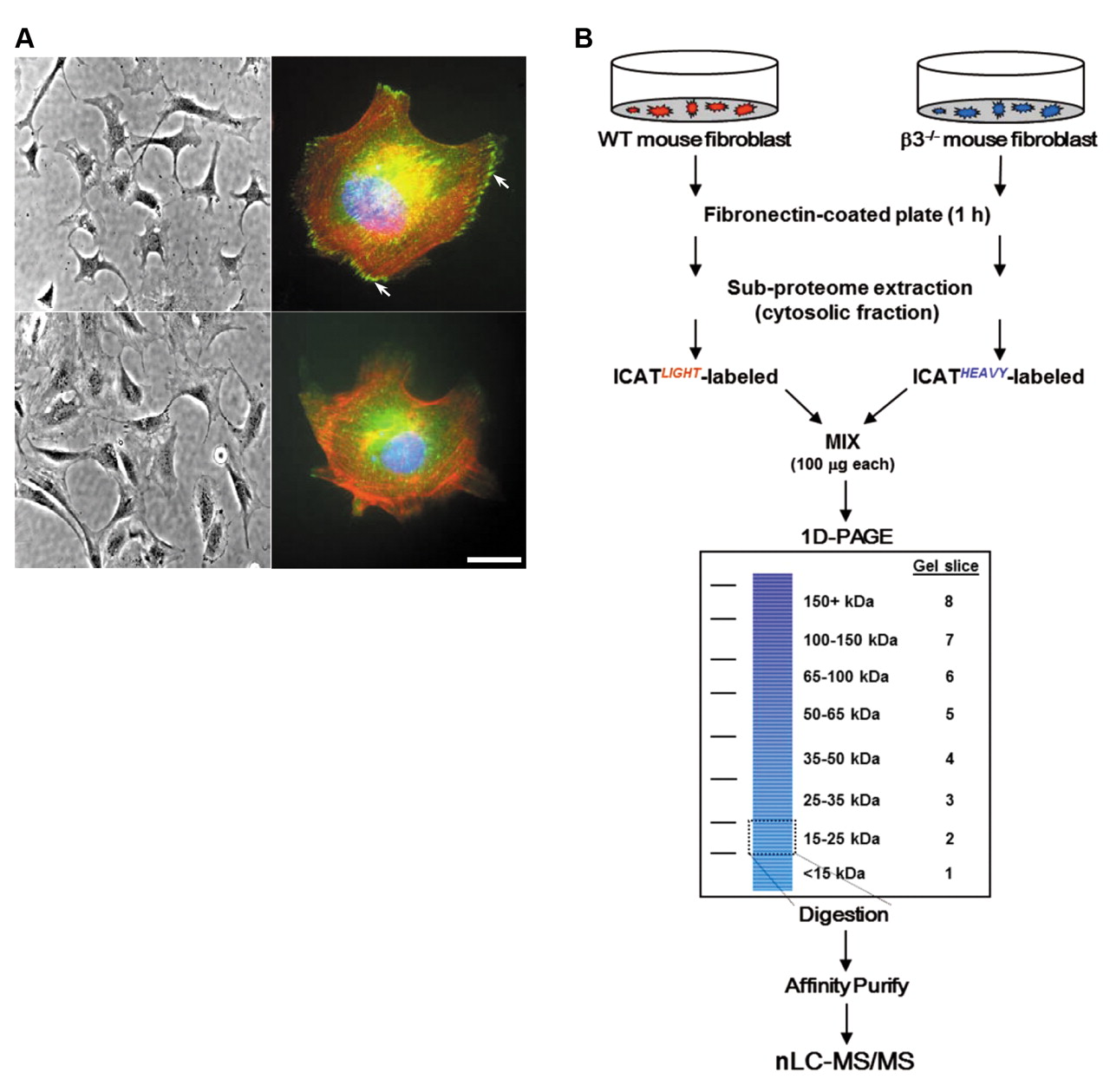
- Figure 1.
Analysis of WT and β3–/– embryonic fibroblasts for quantitative proteomics. A: β3 Integrin deficiency does not change morphology but alters immunofluorescence pattern of paxillin (left panels). Phase-contrast images of WT (top) and β3–/– (bottom) fibroblasts under ×20 magnification (right panels). Merged immunofluorescence images of WT (top) and β3–/– (bottom) fibroblasts stained with anti-paxillin-FITC which localizes to focal contacts (white arrows), phalloidin-Alexa594 (actin), and DAPI (chromatin). Scale bar (bottom right) represents 10 μm. B: Outline of quantitative experimental procedure. Mouse embryonic fibroblasts were seeded onto fibronectin-coated plates, harvested and sub-fractionated. One hundred micrograms of cytosolic protein from WT and β3–/– cells were utilized for ICAT labeling and 1D-PAGE. After Coomassie staining, eight equal-sized gel slices (corresponding to mass ranges of <15, 15-25, 25-35, 35-50, 50-65, 65-100, 100-150, and >150 kDa) were excised and trypsin digested. Digested peptides were further purified over an avidin affinity cartridge. Peptides were eluted from the cartridge then acid-cleaved to release the ICAT-labeled peptides from the linker and subjected to nanoLC-MS/MS analysis. Details of the multidimensional procedure and the nanoLC-MS/MS are described in the Materials and Methods.
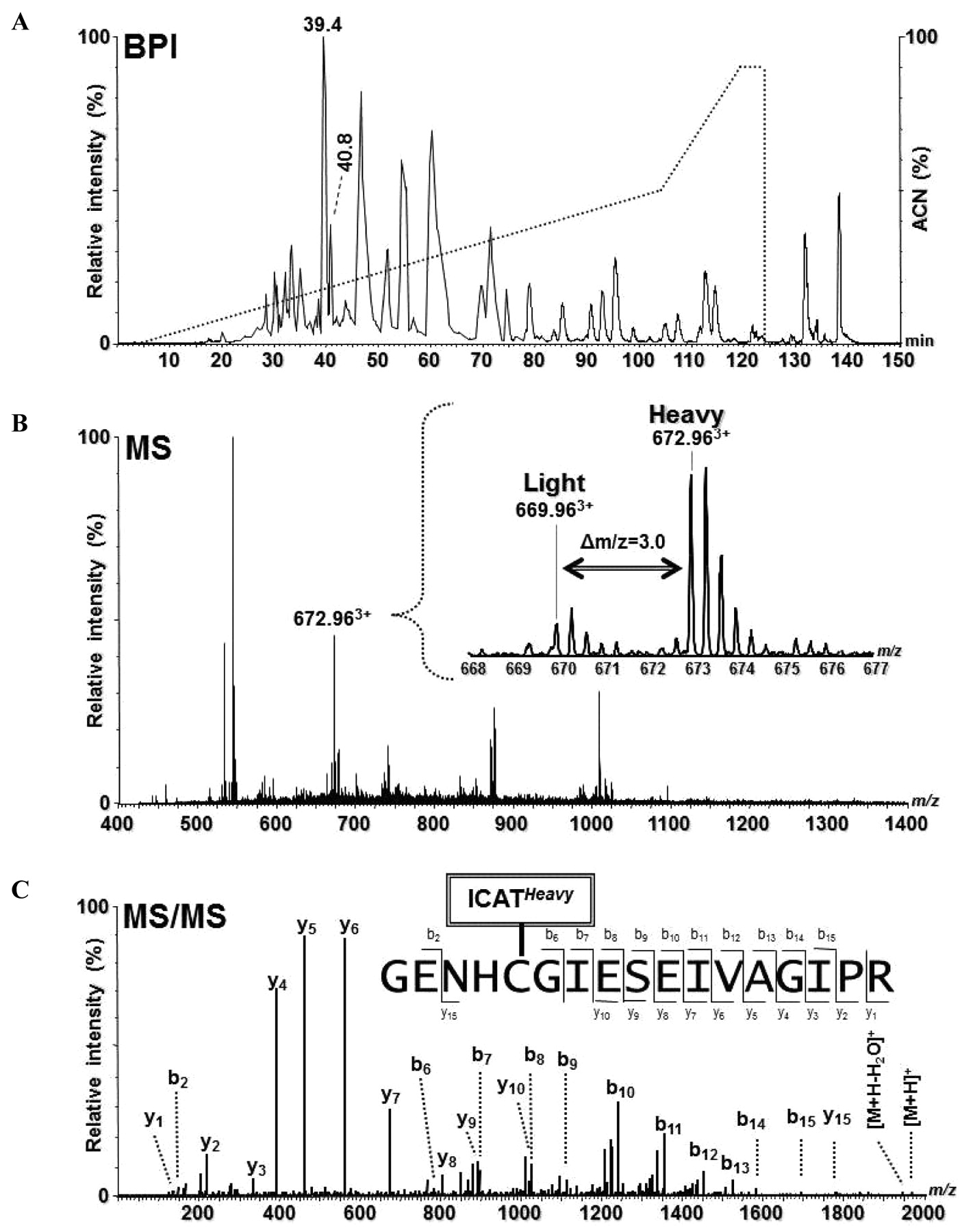
- Figure 2.
Identification and quantitation of CatB of gel slice 4 using ICAT and GeLC-MS/MS. A: Base peak intensity (BPI) chromatogram for nanoLC-MS/MS of ICAT-labeled peptides (solid line) and the organic elution gradient (dotted line) of the acetonitrile (ACN) solvent. B: MS spectrum of the peptides eluting at 40.25 min (between two peaks at retention time 39.4 and 40.8 min in panel A). Inset: Expanded view of the mass spectrum, in which a triple-charged ICAT-labeled peptide pair were observed at m/z 669.96 (light, L) and 672.96 (heavy, H), respectively. H:L ratio=+3.6. C: MS/MS spectrum of the m/z 672.963+ ion resulting from an ICAT-heavy labeled tryptic peptide of CatB from the β3–/– mouse fibroblasts.
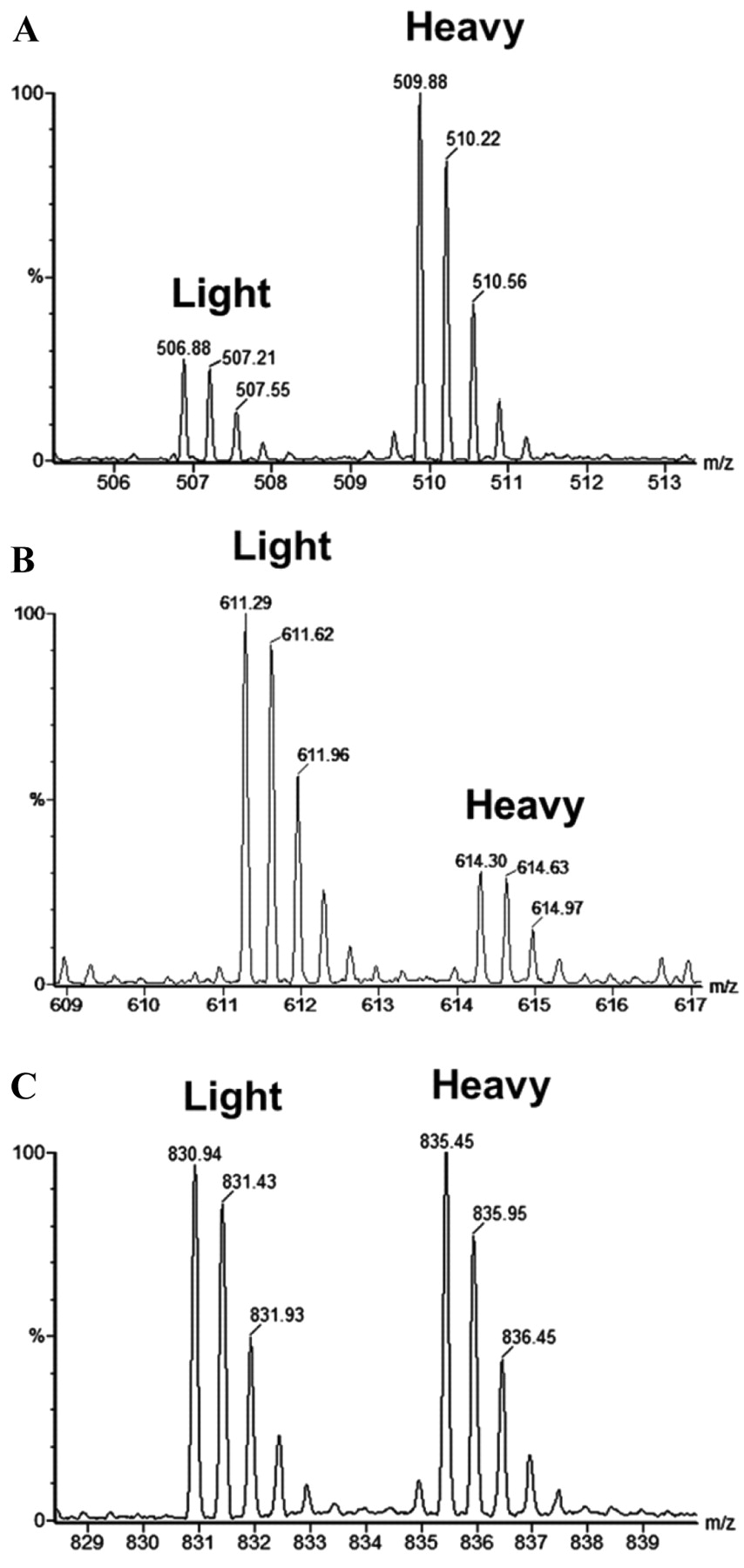
- Figure 3.
Partial MS spectra showing representative pairs of ICAT-light and ICAT-heavy labeled tryptic peptides. A: SEDCFILDHGR from gelsolin, H:L ratio=+2.7. B: FNAHGDANTIVCNTK from galectin-1, H:L=–3.8. C: QVQSLTCEVDALK from vimentin (control), H:L=+1.0. The H(β3–/–):L(WT) ratios were measured automatically using ProteinLynx software, verified by Mascot, and further validated by manual inspection of the extracted ion chromatograms.
- Figure 4.
Independent validation experiments of selected markers from nanoLC-MS/MS experiments of the membrane fraction. A: Immunoblotting correlation against muskelin, cathepsin B, fatty acid binding protein 5, annexin 2, vinculin, galectin-1, and Na+/K+-ATPase (control). B: Numerical values of cICAT quantitation and relative densitometry values from immunoblotting for each pair with WT values equal to 1. C: The log10 values of the western blot densitometry for β3–/– expression were plotted versus the log10 values for cICAT ratios from Tables I and II. Both the slope (y) and the correlation coefficient (R2) of 0.903 were calculated from the represented data.
- Figure 5.
Functional analysis of cathepsin B (CatB) enzymatic activity in cells with reduced or augmented β3 integrin. A fluorometric assay for the determination of CatB proteolytic activity based on cleavage of the synthetic peptide substrate, Z-Arg-Arg-AMC. A: Comparison of cell-associated activity (100 μg protein input) from WT and β3–/– mouse embryonic fibroblast lysates incubated for 1 h with or without the CatB-specific inhibitor, CA-074. B: Activity comparison of secreted pro-CatB from fibroblasts grown on fibronectin in low serum conditions. Media without cells was used as control. C: Comparison of a heterologous cell β3/293 (β3+/+) and HEK293 (β3–/–) human embryonic kidney cell lysates incubated for 1 h with or without the CatB-specific inhibitor, CA-074. Inset: Immunoblot of β3 integrin and CatB protein expression in β3/293 and HEK293. In all experiments, purified recombinant human CatB (10 Units) was used as a control. Values represent the background-subtracted average reading (relative fluorescence units, RFU) from independent experiments repeated at least three times with comparable results.


Tables


In this issue


Jump to section
Related Articles
Cited By...
- No citing articles found.