


Article Figures & Data
Figures
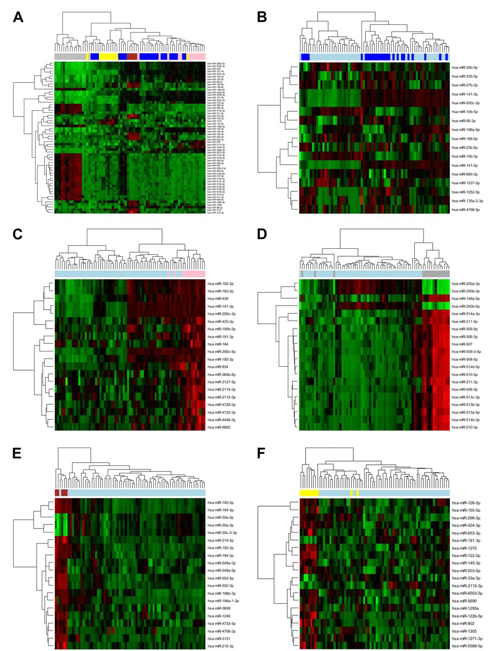
- Figure 1.
Results from differential analysis of miRNA expression assessed by small RNA sequencing in 71 samples of brain metastases (BM) derived from 5 tumor types, including lung carcinomas (dark blue), breast carcinomas (pink), melanomas (gray), colorectal carcinomas (dark red), and renal cell carcinomas (yellow). (A) The miRNA expression profiles in the 5 groups of BMs were compared, resulting in the identification of 58 significantly differentially expressed miRNAs with Benjamini-Hochberg adjusted p-Value smaller than 0.000001. The expression profiles of these miRNAs were visualized in a heatmap with dendrogram. Furthermore, each group was also compared with another group comprised of other BMs. The heatmaps with dendrogram show the results of hierarchical clustering based on the expression of (B) 17 differentially expressed miRNAs (adjusted p-Value <0.05) in BMs derived from lung carcinomas (dark blue) vs. other BMs (light blue), and the 20 most significantly differentially expressed miRNAs in (C) BMs derived from breast carcinomas (pink) vs. other BMs (light blue) (adjusted p-Value ≤0.0017), (D) melanoma-derived BMs (gray) vs. other BMs (light blue) (adjusted p-Value <0.0001), (E) BMs derived from colorectal carcinomas (dark red) vs. other BMs (light blue) (adjusted p-Value ≤0.0002), and (F) BMs derived from renal cell carcinomas (yellow) vs. other BMs (light blue) (adjusted p-Value ≤0.0135). The green-black-red spectrum represents miRNA expression in samples from low (green) to high expression (red).
- Figure 2.
Results from the classifier algorithm and the differential analysis based on the miRNA expression assessed by small RNA sequencing. (A) The classifier algorithm is based on the expression of 6 miRNAs (hsa-miR-141-3p, hsa-miR-141-5p, hsa-miR-146a-5p, hsa-miR-194-5p, hsa-miR-200b-3p, hsa-miR-365b-5p) in 71 samples of BMs derived from lung carcinomas (BML), breast carcinomas (BMB), melanomas (BMM), colorectal carcinomas (BMC), and renal cell carcinomas (BMR). The real BM origin is represented by columns and the classifier prediction by rows. The classifier correctly identified the origin of 91.5% of samples which are highlighted in green. Incorrectly classified samples are highlighted in yellow. The corresponding sensitivity, specificity and 95% confidence interval (CI) values for each group of BMs are shown in the bottom part. (B) The hierarchical clustering analysis based on the expression of the 6 miRNAs in 71 samples of BMs derived from lung carcinomas (dark blue), breast carcinomas (pink), melanomas (gray), colorectal carcinomas (dark red), and renal cell carcinomas (yellow) was visualized in a heatmap with dendrogram. The green-black-red spectrum represents miRNA expression in samples from low (green) to high expression (red).
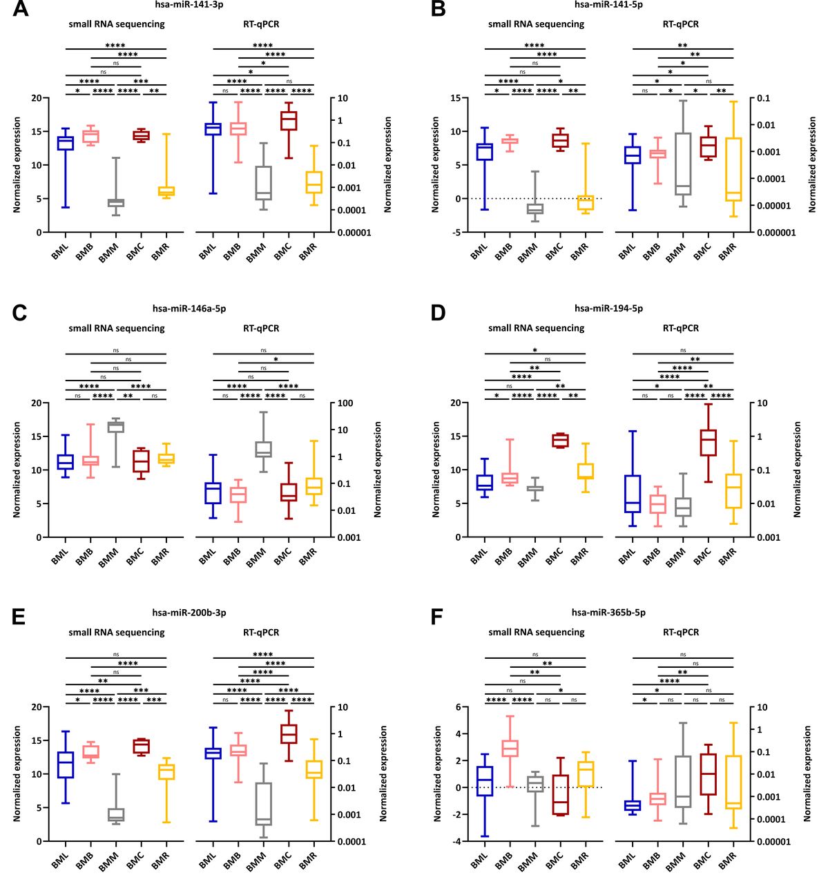
- Figure 3.
Comparison of the expression patterns of the 6 miRNAs; namely (A) hsa-miR-141-3p, (B) hsa-miR-141-5p, (C) hsa-miR-146a-5p, (D) hsa-miR-194-5p, (E) hsa-miR-200b-3p and (F) hsa-miR-365b-5p. The expression was analyzed by small RNA sequencing and RT-qPCR in 71 native tissue samples and 119 formalin-fixed paraffin-embedded tissue samples, respectively, which were derived from lung carcinomas (BML, blue), breast carcinomas (BMB, pink), melanomas (BMM, gray), colorectal carcinomas (BMC, dark red), and renal cell carcinomas (BMR, yellow). Median values of normalized expression were compared using the non-parametric Mann-Whitney U-test. The statistical significance of the results is denoted by ns (not significant; p>0.05), *p<0.05, **p<0.01, ***p<0.001 and ****p<0.0001.
- Figure 4.
Results from the classifier algorithm, which is based on the expression of 6 miRNAs (hsa-miR-141-3p, hsa-miR-141-5p, hsa-miR-146a-5p, hsa-miR-194-5p, hsa-miR-200b-3p, hsa-miR-365b-5p) assessed by RT-qPCR in 119 formalin-fixed paraffin-embedded tissue samples of brain metastases (BM). The real BM origin is represented by columns and the classifier prediction by rows. The classifier correctly identified the origin of 77.3% of samples which are highlighted in green. Incorrectly classified samples are highlighted in yellow. The corresponding sensitivity, specificity and 95% confidence interval (CI) values for each group of BMs are shown in the bottom part.


In this issue


Jump to section
Related Articles
Cited By...
- No citing articles found.