
Main menu

  • Home
  • Current Issue
  • Archive
  • Info for
    • Authors
    • Advertisers
    • Editorial Board
  • Other Publications
    • Anticancer Research
    • In Vivo
    • Cancer Diagnosis & Prognosis
  • More
    • IIAR
    • Conferences
  • About Us
    • General Policy
    • Contact
  • Other Publications
    • Cancer Genomics & Proteomics
    • Anticancer Research
    • In Vivo

User menu

  • Register
  • Subscribe
  • My alerts
  • Log in
  • My Cart

Search

  • Advanced search
Cancer Genomics & Proteomics
  • Other Publications
    • Cancer Genomics & Proteomics
    • Anticancer Research
    • In Vivo
  • Register
  • Subscribe
  • My alerts
  • Log in
  • My Cart
Cancer Genomics & Proteomics

Advanced Search

  • Home
  • Current Issue
  • Archive
  • Info for
    • Authors
    • Advertisers
    • Editorial Board
  • Other Publications
    • Anticancer Research
    • In Vivo
    • Cancer Diagnosis & Prognosis
  • More
    • IIAR
    • Conferences
  • About Us
    • General Policy
    • Contact
  • Visit iiar on Facebook
  • Follow us on Linkedin
Research ArticleArticle
Open Access

Heterogeneity of Platelet Derived Growth Factor Pathway Gene Expression Profile Defines Three Distinct Subgroups of Renal Cell Carcinomas

ADELA MARIA FERICIAN, OVIDIU CATALIN FERICIAN, ANDREI DRAGOS CUMPANAS, PATRICIA LORENA BERZAVA, ALEXANDRU NESIU, ARIANA BARMAYOUN and ANCA MARIA CIMPEAN
Cancer Genomics & Proteomics July 2022, 19 (4) 477-489; DOI: https://doi.org/10.21873/cgp.20334
ADELA MARIA FERICIAN
1Department of Orthopedy and Traumatology/Urology, Victor Babes University of Medicine and Pharmacy, Timisoara, Romania;
  • Find this author on Google Scholar
  • Find this author on PubMed
  • Search for this author on this site
OVIDIU CATALIN FERICIAN
1Department of Orthopedy and Traumatology/Urology, Victor Babes University of Medicine and Pharmacy, Timisoara, Romania;
  • Find this author on Google Scholar
  • Find this author on PubMed
  • Search for this author on this site
  • For correspondence: ovidiu.ferician@yahoo.com ancacimpean1972@yahoo.com acimpeanu@umft.ro
ANDREI DRAGOS CUMPANAS
2Department of Microscopic Morphology/Histology, Victor Babes University of Medicine and Pharmacy, Timisoara, Romania;
3Angiogenesis Research Center, Victor Babes University of Medicine and Pharmacy, Timisoara, Romania;
  • Find this author on Google Scholar
  • Find this author on PubMed
  • Search for this author on this site
PATRICIA LORENA BERZAVA
2Department of Microscopic Morphology/Histology, Victor Babes University of Medicine and Pharmacy, Timisoara, Romania;
3Angiogenesis Research Center, Victor Babes University of Medicine and Pharmacy, Timisoara, Romania;
  • Find this author on Google Scholar
  • Find this author on PubMed
  • Search for this author on this site
ALEXANDRU NESIU
4Department of Urology, Faculty of Medicine “Vasile Goldiş” Western University, Arad, Romania;
  • Find this author on Google Scholar
  • Find this author on PubMed
  • Search for this author on this site
ARIANA BARMAYOUN
5Klinik fur Psychiatrie, Psychosomatik und Psychotherapie, Uniklinikum Frankfurt, Frankfurt, Germany;
  • Find this author on Google Scholar
  • Find this author on PubMed
  • Search for this author on this site
ANCA MARIA CIMPEAN
2Department of Microscopic Morphology/Histology, Victor Babes University of Medicine and Pharmacy, Timisoara, Romania;
3Angiogenesis Research Center, Victor Babes University of Medicine and Pharmacy, Timisoara, Romania;
6Center of Expertise for Rare Vascular Disease in Children, Emergency Hospital for Children Louis Turcanu, Timisoara, Romania
  • Find this author on Google Scholar
  • Find this author on PubMed
  • Search for this author on this site
  • Article
  • Figures & Data
  • Info & Metrics
  • PDF
Loading

Abstract

Background/Aim: We previously described four different vascular patterns (reticular, diffuse, fasciculate, and trabecular) in renal cell carcinoma (RCC) suggesting an early and heterogeneous acquisition of perivascular cells most probably due to a particular PDGF pathway gene expression profile. The aim of the study was to study PDGF pathway gene expression profiles, separately for each vascular pattern. Materials and Methods: TaqMan assay for the PDGF pathway was performed on twelve cases of ccRCC previously evaluated by histopathology, immunohistochemistry, and RNAscope. Gene expression profile was correlated with grade, invasion, vascular patterns, and VEGF. Results: PIK3C3 and SLC9A3 genes were overexpressed in all vascular patterns, but they were significantly correlated with high VEGF mRNA in the reticular and diffuse pattern. STAT1, JAK2, SHC2, SRF and CHUK (IKK) were exclusively overexpressed in cases with diffuse vascular pattern. SLC9A3, CHUK and STAT3 were overexpressed in G2 tumors. Conclusion: Three ccRCC subgroups were defined: 1) PIK3C3 (VSP34)/SLC9A3 which may be proper for anti PIK3C3 inhibitors; 2) VEGFhigh subgroup where association of anti VEGF may be a benefit and 3) JAK2/STAT1 subgroup, potentially being eligible for anti JAK/STAT therapy associated with IKK inhibitors.

Key Words:
  • Renal cell carcinoma (RCC)
  • platelet derived growth factor (PDGF)
  • gene expression profile

Renal cell carcinoma (RCC) represents about 2.2% of total diagnosed cancers worldwide with an estimated number of newly diagnosed patients of 400,000 approximately (1, 2). Fifteen percent of newly diagnosed cancers are diagnosed in the primary metastatic RCC (mRCC) stage of disease but more than 30% of cases which had no metastases at the time of diagnosis become metastatic in a variable period of time of 4 to 8 years from the primary diagnosis despite application of novel and targeted therapies (3). Renal cell carcinoma clear cell type (ccRCC) arises from the proximal tubules of the kidney parenchyma and is the most common subtype of RCC, representing 80% of RCCs (4). ccRCC is a complex disease with a silent development and an unexpected behavior having a highly heterogeneous response to therapy (5). Normal kidney and malignant tissue arising from it have a special and unique microenvironment (6) and this, may be influence the resistance to therapy which appear early during the treatment (7) despite of modern and targeted therapies developed in the last years (8, 9).

Inside renal carcinoma microenvironment heterogeneity, two main components seem to be responsible for the rapid development of therapy resistance: tumor blood vessels and immunogenic profile (10, 11). Vascular and immunogenic compartments of ccRCC are the most “targeted” compartments in ccRCC, antiangiogenic therapies and immune-checkpoints inhibitors being, currently, the gold standard for the treatment of renal cancer.

The heterogeneity of vascular compartment of ccRCC tumor microenvironment is easy to observe also by studying the morphology and immunophenotype of ccRCC tumor vessels. Previously, our team described 4 vascular patterns based on their morphology and cellular components (endothelial cells versus perivascular cells) (12). An unusual tendency to a heterogeneous acquisition of perivascular cells (as time and morphology) was highlighted for tumor blood vessels from ccRCC but also a different arrangement of these vessels inside the tumor. Perivascular cells heterogeneity distribution (assessed by a double immunostaining of tumor blood vessels with CD34/Smooth Muscle Actin antibodies) was the hallmark of the 4 groups of tumor blood vessels from ccRCC defined by our team and was strongly correlated with tumor invasion (12).

Later, in a paper published by Ruis Sauri and colleagues (13) the authors confirmed the presence of the 4 vascular patterns and also reported that these patterns are strongly correlated with ISUP tumor grade (13).

Because of the discrepancies between the high number and unusual morphology of blood vessels from ccRCC (despite of a low endothelial cells proliferation rate) and highly maturated state of these tumor blood vessels, we considered that the study of PDGF-B/PDGFRβ axis is mandatory in ccRCC and correlated with the inhibitory isoform of vascular endothelial growth factor (VEGF), VEGF 165b. In a paper published in 2016 our group reported a significant correlation between VEGF165b and PDGF-B/PDGFRβ ratio (14), this being correlated with vascular patterns previously described. This correlation, exclusively found in the reticular pattern, supports the fact that concomitant inhibition of endothelial cells by VEGF165b and rapid perivascular cell recruitment driven by the PDGF-B/PDGFRβ axis, may define several types of heterogeneous tumor vasculature in ccRCC.

It is well known that any morphology and immunophenotype heterogeneity is supported by a specific gene expression profile (15). Gene expression profiling is a feasible tool to define molecular classes inside various malignancies and based on these stratify prognostic and therapeutic subgroups which may better define the heterogeneous response to therapy, prognosis and survival. The last papers in the field of ccRCC were focused in investigating molecular subgroups of ccRCC (15, 16).

Our previous findings on ccRCC vascular heterogeneity required validation by more specific methods. Tumor blood vessel maturation is given by acquisition of vascular smooth muscle cells driven by PDGF-B/PDGFRβ axis. We aimed to study gene expression profile of the PDGF pathway by using TaqMan™ Array Human PDGF Pathway and correlate the findings with vascular patterns previously described in ccRCC. Further, we aimed to identify potential subgroups of ccRCC able to give a better overview of ccRCC carcinomas with possible different prognostic and therapeutic impact.

Materials and Methods

Case selection. We previously selected 50 retrospective cases of formalin fixed paraffin embedded (FFPE) kidney cancer randomly selected from the archive of Pathology Department of Clinical County Hospital Timişoara, Romania. Biopsies were harvested between 2005 and 2015, fixed in 10% buffered formalin and paraffin embedded following routine protocol. Three independent experienced histopathologists certified microscopic diagnosis of renal cancer, clear cell type. On these cases we previously performed immunohistochemistry, RNAscope and in situ hybridisation focusing on the study of VEGF, PDGF and CD34/SMAct co-expression and we defined four vascular patterns in ccRCC (12). Twelve cases of ccRCC (selected based on vascular patterns previously described by our team (12) were prepared for TaqMan Assay technique. The TaqMan® Array Human PDGF Pathway 96-well plate contains 92 assays to PDGF Pathway associated genes and 4 assays to endogenous control genes. All the experiments were performed in triplicate.

Tissue samples used for RNA extraction were collected by automated image guided coring with Tissue Micro Array technology. Microscopic slides were scanned by using Panoramic Desk Slide Scanner (3Dhistech, Budapest, Hungary) and areas of interest were marked by using automated software of TMA Grand Master (3Dhistech) overlapped to correspondent donor paraffin blocks loaded. Briefly, all 12 donor blocks and correspondent 12 PCR tubes were uploaded into automated TMA web-based digital platform. A 2.0 mm diameter core punch instrument was selected and used to pick up tissue fragments. The specimens are viewed and annotated (marked) using 2.0 mm diameter tool, multiple times using specific color to distinguish tissue areas. Four cores of 2 mm diameter each were collected into 0.2ml PCR tubes from each FFPE block followed by RNA extraction using a semi-automated method described below.

RNA extraction by semi-automated magnetic beads-based technology and cDNA synthesis. Dewaxing of FFPE tissue cores was performed by filling PCR tubes containing material with xylene and incubating them for 60 minutes at room temperature for cores dewaxing. Xylene was discarded and RNA extraction was started by using King Fisher Pure RNA Tissue Kit (Thermo Scientific, Waltham, MA, USA). Reagent preparation was done according to the protocol provided by the manufacturer and loaded into Kingfisher 96 Deep Well Plates based on the predesigned template included in the protocol. RNA extraction continued by loading the plates into King Fisher Duo Prime automated System (Thermo Scientific, Santa Clara, CA, USA) and by choosing Pure RNA Tissue Duo program as running protocol. The RNA total amount was checked using a Qubit™ 3.0 fluorometer (Thermo Scientific, Santa Clara, USA) by preparing the preliminary samples with the Qubit™ RNA HS Analysis Kit (code Q32852, Thermo Scientific). cDNA synthesis from the extracted RNA was performed by using the Applied Biosystem High-capacity RNA-to-cDNA kit™ (Fischer Scientific, Loughborough, UK) following the protocol provided by the manufacturer. Two micrograms of total RNA were used per 20-μl reaction. The TaqMan Array plates were prepared by loading 20 μl of cDNA sample prepared by using 10 μl of nuclease-free water and 10 μl of Master Mix (2x) on each well. The TaqMan Array Human PDGF plates were subjected of RT-PCR technique performed on the 7500 Fast Dx Real-Time PCR Instrument (Agilent, Santa Clara, CA, USA).

Immunohistochemistry. The cases for TaqMan Array were selected based on previous assessments of immunohistochemical stained slides with a double-stain method for endothelial cells (monoclonal mouse anti-human CD34, clone QBEnd 10, Novocastra, Newcastle upon Tyne, UK) and for perivascular cells (monoclonal mouse anti-human Smooth Muscle Actin antibody, clone 1A4, Novocastra). Each step of immunohistochemical procedure was done in an automated manner by using software of Bond Max Autostainer (Leica MicroSystems, Deer Park, IL, USA). Thirty minutes incubation time with primary antibodies was followed by the use of Bond Refine Detection System Dual Color (Leica MicroSystems). Based on the CD34/SMAct coexpression we selected cases according to our team classification reported by Ferician et al. (15) as we include all four types of vascular patterns previously described.

Data analysis. The microscopic assessment of CD34/SMAct was performed by three independent pathologists using AxioZoom Zeiss Microscope (Zeiss, Oberkochen, Germany). Interaction between CD34 positive endothelial cells and SMAct positive perivascular cells was the main criterion used to select cases for TaqMan Array. TaqMan assay results were evaluated by using Data Assist V.3.01 software (Thermo Fisher Scientific, Waltham, MA, USA). Heat Map and volcano plots were automatically generated from this analysis.

Results

We previously characterized all 12 cases of ccRCC included in the present study regarding tumor grade, vascular pattern, the presence of CD34/SMAct immunoexpression, and assessment of tumor blood vessels stabilization (a parameter which make ccRCC less responsive to antiangiogenic and/or antivascular therapy). Initial microscopic assessment of each ccRCC specimen included the evaluation of tumor grade. Eight cases were scored as having tumor grade 2 and 4 cases with tumor grade 3. We detected a high angiogenic process inside tumor mass (Figure 1) with a high density of tumor blood vessels being in all stages of angiogenic process (buds, cords, tube-like structures, perfused vessels). Despite an active angiogenic morphological appearance, even for angiogenic stages where SMAct-positive perivascular cells should be absent (buds and cords stages), we observed SMAct-positive perivascular cells with a heterogeneous distribution.

Figure 1.
  • Download figure
  • Open in new tab
  • Download powerpoint
Figure 1.

Intense tumor angiogenic process inside renal cell carcinoma clear cell type (ccRCC). Note tumor blood vessels heterogeneity inside the same microscopic field, containing vascular buds (sign of an intense endothelial cells activation), cords (suggesting an active endothelial cells migration and small vascular structures with lumen (as already functional tumor blood vessels). Yellow arrows indicate early presence of smooth muscle actin (SMAct) positive perivascular cells which usually appear only in stabilisation stage of tumor blood vessels. As herein shown, perivascular cells are partially attached to the outer part of the vessel, some of them having unusual extensions in between two newly formed blood vessels (yellow arrows, lower part of the image).

All cases showed stable tumor blood vessels highlighted by CD34/SMAct immunostaining in the whole area or just in spotted zones of tumor mass (Figure 2a). Most tumor blood vessels showed an oversized perivascular cell network highlighted with smooth muscle actin (SMA) immunostaining (Figure 2b). Also, the arrangement of SMA-positive perivascular cells was not normal (closely attached to the outer circumference of the vessel wall), being partially detached (Figure 2b) or being distributed as bridge-like structures in between small tumor blood vessels loops (Figure 2b).

Figure 2.
  • Download figure
  • Open in new tab
  • Download powerpoint
Figure 2.

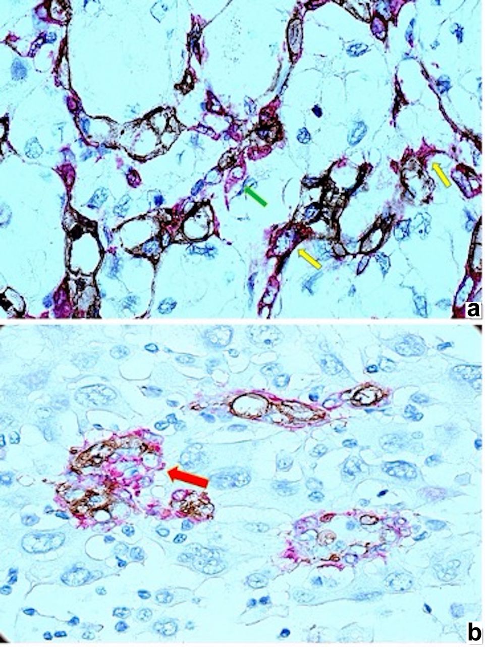
Early presence, particular morphology and distribution of smooth muscle actin (SMAct) positive perivascular cells related to renal cell carcinoma clear cell type (ccRCC) tumor blood vessels. Incomplete attachment of perivascular cells on tumor blood vessels [(a), green arrow, stained in red)] and bridge-like arrangement of smooth muscle actin (SMAct) positive perivascular cells (stained in red) in between tumor vessels [(a), yellow arrows)]. Other tumor areas have glomeruloid like structures [(b), red arrow)] with smooth muscle actin (SMAct) positive perivascular cells surrounding CD34 positive structures without evident lumen (most probably newly formed blood vessels in early stages of angiogenesis).

Based on these preliminary microscopic observations together with a high immunohistochemical expression of PDGF-BB previously reported by our team (17) and given the fact that perivascular cells acquisition is driven by PDGF-B/PDGFRβ pathway, we performed TaqMan Array to highlight the peculiarities of PDGF pathway gene expression to identify potential gene overexpression related to unusual early presence, morphology and distribution of SMAct-positive perivascular cells.

Immunohistochemical evaluation of PDGF-B/PDGFRβ showed that all 12 cases of ccRCC were positive for PDGF-BB, by immunohistochemistry and RNAscope of which 91.6% were confirmed by RT-PCR.

PDGF pathway gene expression profile was evaluated related to our previous results regarding VEGF immunohistochemical and RNAscope expression and also with our previous classification which stratifies the types of tumor blood vessels from ccRCC in 4 categories: reticular, diffuse, fascicular, trabecular.

Based on TaqMan Array results analysis, a PDGF pathway gene expression profile HeatMap has been automated created by Data Assist analysis software. As it has been shown in Figure 3, HeatMap analysis revealed several overexpressed PDGF pathways genes. Genes with the highest overexpression were: PIK3C3 (VsP34), SLC9A3, STAT1, JAK2, SHC2, SRF and CHUK.

Figure 3.
  • Download figure
  • Open in new tab
  • Download powerpoint
Figure 3.

Heat Map of platelet derived growth factor (PDGF) pathway automatically generated based on TaqMan Assay analysis. Gene overexpression was highlighted in red, gene down-regulation in green and the lack of expression in black. As shown in the heatmap, overexpressed genes were: PIK3C3 (VsP34), SLC9A3, STAT1, JAK2, SHC2, SRF and CHUK.

Volcano plot analysis was correlated with ccRCC vascular patterns, VEGF immunohistochemical expression and tumor grade.

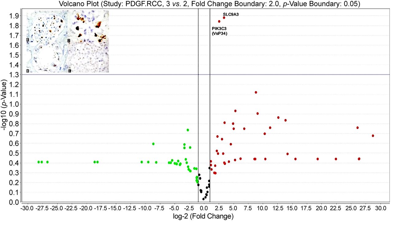
PIK3C3 (VsP34) and SLC9A3 were up-regulated (Figure 4) for both reticular and diffuse vascular patterns ccRCC cases which had a moderate and intense VEGF immunohistochemical expression (VEGF score 2 ore 3) certified by RNAscope in situ hybridization method (Figure 4, inset).

Figure 4.
  • Download figure
  • Open in new tab
  • Download powerpoint
Figure 4.

Volcano plot showing statistically significant up-regulated genes: SLC9A3 and PIK3C3, common for reticular and diffuse vascular patterns of ccRCC.

In the reticular vascular pattern ccRCC group, vascular invasion was found in 66.6% out of the total number of cases. For this group, PDGF pathway TaqMan analysis revealed a statistically significant MAP2K2 up-regulation (p=0.004). For the diffuse and trabecular pattern group we detected vascular invasion for 40% of cases. Analysis of PDGF pathway gene expression profile identified other two up-regulated genes specific for this group: PIK3C3 (p=0.004211) and MAPK3 (p=0.048) (Figure 5). MAPK3 was found to be up-regulated also in group with high expression of VEGF inhibitory isoform, VEGF 165b (p=0.0127).

Figure 5.
  • Download figure
  • Open in new tab
  • Download powerpoint
Figure 5.

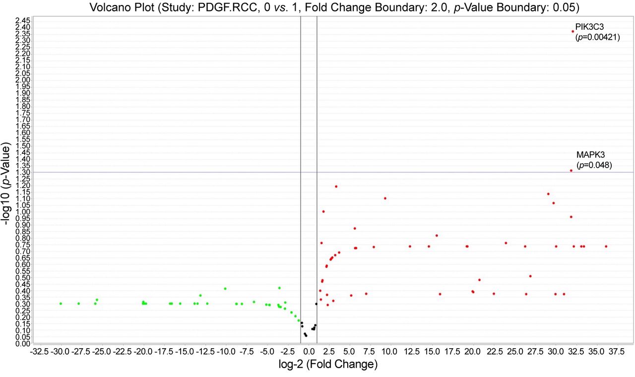
Volcano plot showing gene up-regulation for diffuse vascular pattern cases with invasion.

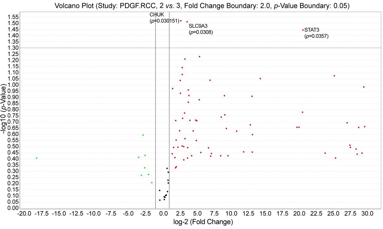
PDGF pathway TaqMan assay was also assessed related to tumour grade. CHUK (p=0.030151), STAT3 (p=0.0357) and SLC9A3 (p=0.0308) genes were statistically significant up-regulated for ccRCC cases having tumor grade 2 (see volcano plot from Figure 6).

Figure 6.
  • Download figure
  • Open in new tab
  • Download powerpoint
Figure 6.

Platelet derived growth factor (PDGF) pathway gene expression profile assessment related to tumor grade. Grade 2 was characterized by significant up-regulation for 3 genes compared to other tumor grades.

Five new different genes have been found to be down-regulated in ccRCC cases with reticular pattern (Figure 7) and up-regulated for diffuse pattern exclusively. These genes are: STAT1, JAK2, SHC2, SRF, CHUK.

Figure 7.
  • Download figure
  • Open in new tab
  • Download powerpoint
Figure 7.

STAT1, JAK2, SHC2, SRF, CHUK down-regulation for the diffuse and reticular pattern.

Based on gene analysis of the PDGF pathway in ccRCC correlated with VEGF expression and tumor blood vessels types, we defined three subgroups of renal carcinomas which may have three different therapeutic options. The first is the VEGFhigh subgroup showing PIK3C3 (VPS34) and SLC9A3 up-regulation. This group includes both reticular and diffuse vascular patterns and might be eligible to a combined therapeutic option of anti-VEGF agents and PIK3C3 inhibitors. The second group may be defined as VEGFlow/- showing a similar PIK3C3 (VPS34), SLC9A3 up-regulation. For this group, anti-PDGF-PDGFR may be suitable. The third subgroup that we called JAK/STAT subgroup is characterized by overexpression of the 5 genes, suitable for anti JAK/STAT therapy associated with PIK3C3 and IKK inhibitors (Figure 8).

Figure 8.
  • Download figure
  • Open in new tab
  • Download powerpoint
Figure 8.

Proposed renal cell carcinoma clear cell type (ccRCC) subgroups based on Platelet Derived Growth Factor (PDGF) pathway assessment correlated to ccRCC tumour vascular pattern.

Discussion

Renal cancer remains one of the most unpredictable malignancies related to its intratumor heterogeneity, invasiveness, metastatic behaviour, response to conventional and targeted therapies (17, 18) as well as to high vascularisation and heterogeneity of tumour blood vessels (12, 19). VHL gene mutation has been reported for several years as one of the main inductors of ccRCC initiation and progression, while other gene expression profile pathways (as angiogenic or lymphangiogenic pathways gene expression profile) being somehow neglected for several years.

Most of ccRCC are characterized by an increased density of intratumor blood vessels at the time of diagnosis. Intratumor blood vessels have a highly heterogeneous morphology previously described and classified by our team in four vascular patterns based on their microscopic morphology, presence and arrangement of perivascular cells (12). Three out of four vascular patterns described before (12) included tumor blood vessels showing a high grade of maturation certified by the presence of SMA-positive perivascular cells. Following our paper related to vascular pattern heterogeneity (12), other reports continued to be published being increasingly focused on ccRCC tumor blood vessels heterogeneity and its potential impact on the development of resistance to targeted therapies (13, 20-22).

Relatively recent data suggested that there is a strong mutual communication in between tumor cells and intratumor blood vessels. The relationship between the tumor microenvironment and ccRCC tumor cell heterogeneity is increasingly reported in the literature both on experimental models, and by gene expression analysis of human tissues derived from patients with renal cancer (23, 24). Intratumor and peritumour microenvironment is highly heterogeneous not only by its cellular components, such as tumor fibroblasts (25) or macrophages (26), but also by its vascularisation (12). Yuan and colleagues, reported in 2016 that highly perfused ccRCC tumor areas, have increased cellularity and increased microvessel density compared to low-perfused areas. This suggests a direct correlation between vascularity and tumor cells in ccRCC (27). Following this report, one of the first to suggest the interrelation between tumor cell behavior and renal tumor vascularization, several studies have confirmed that tumor cell aggressiveness and their response to therapy are closely related to tumor vascularization (27-30). As evidence of the essential involvement of blood vessels in the behavior of tumor cells, most targeted therapies used in the treatment of ccRCC, target tyrosine kinases and their receptors (31, 32). Under these targeted therapies recurrence and metastasis rates decreased significantly but resistance to therapy still developed by incompletely elucidated mechanisms (33, 34).

Despite the above, there are few studies that refer to the morphology of blood vessels, as well as the involvement of perivascular cells in the early maturation of ccRCC tumor vessels. Ferician and colleagues, classified intratumoral blood vessels into 4 subtypes depending on the presence and architecture of perivascular cells (12). The same team showed that VEGF165b has a major contribution in the phosphorylation of PDGF-beta. This combination most likely cause inhibitory effects on endothelial proliferation and migration, which partly explains the early maturation of blood vessels with the 4 different patterns described above.

Morphological and immunohistochemical ccRCC tumor blood vessels heterogeneity certified by previous mentioned studies suggested a possible PDGF pathway gene expression profile heterogeneity which was the aim of the present study.

TaqMan analysis revealed two genes overexpression (PIK3C3 (VPS34) and SLC9A3) common to ccRCC diffuse and reticular vascular patterns for cases with moderate and high VEGF expression.

PIK3C3 also called VPS34, is involved in intracellular membrane trafficking and autophagy, and is a target for a SAR405 compound approved in 2014 together with Everolimus, the combination giving synergistic effects as an antiproliferative of renal cancer tumor cells (35). In 2020 Jones and colleagues described three VPS34 inhibitors with proved effects on ccRCC tumor cell autophagy (36).

Although there are numerous studies on autophagy of tumor cells, studies related to autophagy in smooth muscle cells and endothelial cells of tumour blood vessels normalization are limited (37).

Currently, there are studies proving that overexpression of the PIK3C3 gene (VPs34) stimulates proliferation of perivascular smooth muscle cells in the experimental model by differentiating their SUMO1-dependent, as a morphological substrate responsible for pulmonary arterial hypertension (38). These data, correlated with the microscopic observations of an early maturation of the newly recruited tumor blood vessels in ccRCC as well as the gene overexpression of PIK3C3 (VPs34), reported in our study, support the hypothesis that this gene is not only involved in the autophagy mechanism of tumor cells but also acts on perivascular smooth muscle cells autophagy, practically performing an early remodeling of tumor blood vessels.

This may partially explain the early maturation of ccRCC tumor blood vessels, most of these tumours already having tumor blood vessels covered by perivascular cells at the time of diagnosis (20). Early maturation of tumor vessels results in a partial response or a development over time of partial or total resistance to antiangiogenic and/or antivascular therapies actually used in ccRCC.

If for perivascular smooth muscle cells the role of PIK3C3 (VPs34) is partially explained and accepted in benign lesions and less in malignant ones, the involvement of PIK3C3 (VPs34) in endothelial cell activation seems to be closely correlated with VEGF overexpression. The performance of this function is related to the interaction with the caveolin system in the endothelial cell membrane (39).

The role of PIK3C3 (VPs34) in tumor angiogenesis is indirectly suggested at this time by its interaction with the VEGF pathway, but also with hypoxia in the tumor microenvironment mediated by the HIF1α pathway (40). A proposed mechanism for the involvement of PIK3C3 (VPs34) in tumor angiogenesis is that of interaction with tyrosine kinase receptors, forming currently important targets in ccRCC therapy (41).

PIK3C3 (VPs34) is involved in the cellular internalization of VEGFR2 (41). This aspect, correlated with the maturation of blood vessels, may partly explain the low rate of endothelial cell proliferation in tumor vessels despite VEGF overexpression in tumor cells described by our team in previous studies (42).

SLC9A3, the second overexpressed gene in our study, in cases with diffuse and reticular vascular network and increased VEGF is known and studied as an ion transporter in the renal parenchyma (43). Its action on vascular smooth muscle cells is scarcely studied in the literature. The experimental model of pulmonary hypertension in rats demonstrated that overexpression of the NHE family of which SLC9A3 is a part causes proliferation and migration of vascular smooth muscle cells that are responsible for pulmonary hypertension (44, 45). The role of this gene in tumor angiogenesis is unknown. Its overexpression in our study may be related either to a study published by Karumanchi et al. (46) where the authors reported its overexpression in renal cell carcinoma cells dependent on the von Hippel-Lindau gene mutation (VHL) so common in renal cell carcinoma, or an action of hyperplasia of vascular smooth muscle cells that causes early maturation of tumor vessels in the ccRCC.

For the diffuse pattern of ccRCC vascularization, we identified the overexpression of 5 genes, most of them being part of the JAK/STAT pathway well known as having a major role in tumor angiogenesis. Activation of the JAK/STAT pathway causes proliferation of perivascular smooth muscle cells, with their hyperplasia, especially the JAK2/STAT3 axis (47) and JAK2/STAT1 (48) through a PDGF-dependent mechanism.

The JAK/STAT pathway is intensely studied in renal cell carcinoma progression and metastasis (49-51) as well as in ccRCC-associated inflammation (52) but the association between this pathway and tumor blood vessel types has not been performed to date. We found that JAK2 and STAT1 were up-regulated in our study related to ccRCC group having a diffuse pattern of tumor blood vessels which showed a high maturation grade by the presence of perivascular smooth muscle cells. Recently, Kong et al. (53) reported that the JAK2/STAT1 pathway is correlated with PD-L1 activation and expression mediated by HIF2α and IFNγ. HIF2α up-regulates genes involved in tumor blood vessel maturation (53). The JAK2/STAT1 pathway has been also reported to be involved in vascular remodeling by acting on vascular smooth muscle cells during different steps of atherosclerosis or following vascular injuries (54, 55). Bartoli et al. demonstrated that VEGF activates STAT1 in bovine aortic endothelial cells by facilitating the interaction between STAT1 and VEGFR2 (56). This may explain our finding of JAK2/STAT1 genes up-regulation in ccRCC cases with moderate and intense expression of VEGF from both diffuse and reticular pattern.

The origin of smooth muscle perivascular cells obtained by ccRCC is currently unknown. Also, the molecular mechanisms involved in the early and rapid acquisition of perivascular smooth muscle cells around ccRCC tumor vessels are unknown. Normal kidney contains cells (in both parenchyma and stroma) with the ability to transdifferentiate in smooth muscle-like cells which might be recruited by ccRCC tumor vessels during their maturation. Mesangial cells (considered to be modified pericytes) (57) were recently classified by He et al. (58) as having two distinct phenotypes: a pericyte-like phenotype and a fibroblast-like phenotype. Also, mesangial cells express FoxD1 gene which is also expressed by a population of progenitor cells that give rise to renal stroma, pericytes, vascular smooth muscle cells and mesangial cells (59-61).

The JAK2/STAT1 pathway is activated in mesangial cells during kidney response to an injury as diabetic nephropathy (62) and renal fibrosis (63) but their involvement in kidney malignancies is not yet reported. JAK2/STAT1 pathway is controlled by PDGF (62). In the normal human kidney, a high PDGF expression was reported inside mesangial cells which also have PDGF receptors (63). Moreover, PDGF has an autocrine effect on mesangial cells inducing their proliferation (64). During embryonic development of renal glomerulus, PDGF secreted by endothelial progenitor cells attracts mesangial cells and stimulates their attachment to the outer side of the capillaries inducing glomerular capillaries loops development. Our microscopic assessment of smooth muscle cells attached to ccRCC tumor blood vessels linked to our molecular findings and also related to previous presented indirect evidence suggests mesangial cells as a potential source of tumor blood vessels perivascular cells by a mechanism mediated by PDGF/PDGFRs activation through JAK2/STAT1 pathway. In our study, all 12 cases were PDGF positive by RNAscope and immunohistochemistry and in more than 90% this expression was confirmed by RT PCR. Simon et al. demonstrated that PDGF has a mitogenic effect on human airways smooth muscle cells and this effect is mediated in part by JAK2/STAT1 pathway (48). In our study the diffuse pattern of tumor vasculature includes PDGF-positive ccRCC cases having intratumor blood vessels with a continuous layer of SMAct positive perivascular cells. For this group JAK2 and STAT1 up-regulation found by TaqMan analysis may explain, in part, the rapid maturation of tumor blood vessels most probably due to mitogenic effects of PDGF on perivascular smooth muscle cells by a mechanism involving JAK2 and STAT1. PDGF stimulated smooth muscle cells activates STAT1 alpha (p91) (65) and switch to a secretory phenotype for growth factors and IFN γ (65).

STAT1 activation decreased expression of proangiogenic molecules bFGF, MMP-2, and MMP-9 and subsequently may induce a decrease of endothelial cells activation (66, 67). STAT1 action on both endothelial and perivascular cells may explain the presence of already mature tumor blood vessels at the time of ccRCC diagnosis (67).

STAT1 increased in radioresistant human ccRCC samples and inhibition of STAT1 by fludarabine and siRNAs enhanced ccRCC cell radiosensitivity (67). Banes-Berceli et al. (68) reported that levels of JAK1 and JAK2 mRNA were higher in ccRCC cells compared to normal kidney tissue and this molecular mechanism may be responsible for inappropriate cell survival and chemoresistance. Sunitinib therapy influences JAK2/STAT1 pathway by magnifying IFNg-induced STAT1 activity and causing an increase of the activity of janus kinase 2 (JAK2) (69). The same authors linked STAT1 up-regulation by indoleamine 2,3-dioxygenase (IDO) activation.

IDO has been reported to be expressed in mature tumor associated blood vessels from early stage cervical cancer (70). Also, IDO is known to be one of the immune checkpoint regulators (71). Recently, Jonasch et al. (72) proposed combination of anti-angiogenics and checkpoint inhibitors for renal cell carcinoma as a novel therapeutic strategy based on promising results of preclinical studies showing the benefits of such association, some of them targeting perivascular cells detachments. Our finding about JAK2/STAT1 pathway up-regulation for ccRCC group with diffuse pattern of tumor blood vessels may sustain this novel therapeutic strategy.

Serum response factor (SRF) was also up-regulated in diffuse vascular pattern ccRCC group. SRF increases ccRCC tumor cells migration and invasion by stimulating epithelial to mesenchymal transition (73). SRF gene is well known to have a dual role on both endothelial cells and perivascular cells. SRF is responsible for actin cytoskeleton dynamics and is critical for EC filopodia formation, tip cell contractility and EC migration (74, 75). Also, SRF is involved in epithelial to pericyte transition reported in renal tubular epithelial cells (76) as well as in cancer cells (77). This SRF action on both endothelial and perivascular cells may explain in part the high microvessels density in ccRCC, but also the predominance of mature type tumor blood vessels.

Conclusion

We herein demonstrated PDGF pathway gene expression profile heterogeneity of ccRCC related to vascular tumor pattern, invasion status and tumor grade. By correlating PDGF pathway gene expression profile and ccRCC vascular patterns we defined 3 ccRCC distinct subgroups which may have a potential impact on choosing the best therapeutic strategy for each patient. Further studies are needed to elucidate ccRCC tumor blood vessels perivascular cell origin and also to assess molecular mechanisms responsible for the early and rapid maturation of ccRCC tumor blood vessels.

Acknowledgements

The Authors are grateful to Ciprian Onica and Raluca Amalia Ceausu for their great technical support and also to Pathology Department of County Hospital Timisoara for providing us FFPE blocks. All our gratitude to Victor Babes University of Medicine and Pharmacy Timisoara, Romania for its continuous financial support for covering publishing fees and for the logistics.

Footnotes

  • Authors’ Contributions

    AMF, OCF and AN obtained ccRCC tumours sample, FFPE blocks and selected them for immunohistochemistry and molecular studies. AMF and OCF designed the study and wrote the paper. ADC, PLB and AB (former student of VADA program from Histology Department, currently PhD student at Uniklinikum Frankfurt) performed all steps of molecular analysis. AMC supervised the study by validating each preliminary and final results and also reviewed the final draft.

  • Conflicts of Interest

    The Authors have no conflicts of interest to declare.

  • Received March 18, 2022.
  • Revision received April 18, 2022.
  • Accepted May 11, 2022.
  • Copyright © 2022, International Institute of Anticancer Research (Dr. George J. Delinasios), All rights reserved

This article is an open access article distributed under the terms and conditions of the Creative Commons Attribution (CC BY-NC-ND) 4.0 international license (https://creativecommons.org/licenses/by-nc-nd/4.0).

References

  1. ↵
    Global Cancer Observatory: Cancer Today. Lyon, France, International Agency for Research on Cancer. Available at: https://gco.iarc.fr/today [Last accessed on March 2, 2020]
  2. ↵
    1. Padala SA,
    2. Barsouk A,
    3. Thandra KC,
    4. Saginala K,
    5. Mohammed A,
    6. Vakiti A,
    7. Rawla P and
    8. Barsouk A
    : Epidemiology of renal cell carcinoma. World J Oncol 11(3): 79-87, 2020. PMID: 32494314. DOI: 10.14740/wjon1279
    OpenUrlCrossRefPubMed
  3. ↵
    1. Li C,
    2. Wang R,
    3. Ma W,
    4. Liu S and
    5. Yao X
    : Do metastatic kidney cancer patients benefit from cytoreductive nephrectomy? A real-world retrospective study from the SEER database. Front Surg 8: 716455, 2021. PMID: 34557516. DOI: 10.3389/fsurg.2021.716455
    OpenUrlCrossRefPubMed
  4. ↵
    1. Athanazio DA,
    2. Amorim LS,
    3. da Cunha IW,
    4. Leite KR,
    5. da Paz AR,
    6. de Paula Xavier Gomes R,
    7. Tavora FRF,
    8. Faraj SF,
    9. Cavalcanti MC and
    10. Bezerra SM
    : Classification of renal cell tumors - current concepts and use of ancillary tests: recommendations of the Brazilian Society of Pathology. Surgical and Experimental Pathology 4(1): 4, 2021. DOI: 10.1186/s42047-020-00084-x
    OpenUrlCrossRef
  5. ↵
    1. Hsieh JJ,
    2. Purdue MP,
    3. Signoretti S,
    4. Swanton C,
    5. Albiges L,
    6. Schmidinger M,
    7. Heng DY,
    8. Larkin J and
    9. Ficarra V
    : Renal cell carcinoma. Nat Rev Dis Primers 3: 17009, 2017. PMID: 28276433. DOI: 10.1038/nrdp.2017.9
    OpenUrlCrossRefPubMed
  6. ↵
    1. Chen W,
    2. Pan X and
    3. Cui X
    : RCC immune microenvironment subsequent to targeted therapy: a friend or a foe? Front Oncol 10: 573690, 2020. PMID: 33117708. DOI: 10.3389/fonc.2020.573690
    OpenUrlCrossRefPubMed
  7. ↵
    1. Makhov P,
    2. Joshi S,
    3. Ghatalia P,
    4. Kutikov A,
    5. Uzzo RG and
    6. Kolenko VM
    : Resistance to systemic therapies in clear cell renal cell carcinoma: mechanisms and management strategies. Mol Cancer Ther 17(7): 1355-1364, 2018. PMID: 29967214. DOI: 10.1158/1535-7163.MCT-17-1299
    OpenUrlAbstract/FREE Full Text
  8. ↵
    1. Mikami S,
    2. Mizuno R,
    3. Kosaka T,
    4. Tanaka N,
    5. Kuroda N,
    6. Nagashima Y,
    7. Okada Y and
    8. Oya M
    : Significance of tumor microenvironment in acquiring resistance to vascular endothelial growth factortyrosine kinase inhibitor and recent advance of systemic treatment of clear cell renal cell carcinoma. Pathol Int 70(10): 712-723, 2020. PMID: 32652869. DOI: 10.1111/pin.12984
    OpenUrlCrossRefPubMed
  9. ↵
    1. Sharma R,
    2. Kadife E,
    3. Myers M,
    4. Kannourakis G,
    5. Prithviraj P and
    6. Ahmed N
    : Determinants of resistance to VEGF-TKI and immune checkpoint inhibitors in metastatic renal cell carcinoma. J Exp Clin Cancer Res 40(1): 186, 2021. PMID: 34099013. DOI: 10.1186/s13046-021-01961-3
    OpenUrlCrossRefPubMed
  10. ↵
    1. Canete-Portillo S,
    2. Rodriguez Pena MDC,
    3. Wang D,
    4. Sanchez DF,
    5. Netto GJ and
    6. Magi-Galluzzi C
    : Vascular architectural patterns in clear cell renal cell carcinoma and clear cell papillary renal cell carcinoma. Virchows Arch 479(6): 1187-1196, 2021. PMID: 34611731. DOI: 10.1007/s00428-021-03214-2
    OpenUrlCrossRefPubMed
  11. ↵
    1. Liu S,
    2. Xu C,
    3. Zhang K,
    4. Han D,
    5. Yang F,
    6. Li Y,
    7. Zhao X,
    8. Ma S,
    9. Li H,
    10. Lu S,
    11. Lu T,
    12. Zhang J,
    13. Qin W,
    14. Wen W and
    15. Yang B
    : CD248 as a bridge between angiogenesis and immunosuppression: a promising prognostic and therapeutic target for renal cell carcinoma. Ann Transl Med 9(23): 1741, 2021. PMID: 35071435. DOI: 10.21037/atm-21-6271
    OpenUrlCrossRefPubMed
  12. ↵
    1. Ferician O,
    2. Cimpean AM,
    3. Ceausu AR,
    4. Dema A,
    5. Raica M and
    6. Cumpanas A
    : Heterogeneous vascular patterns in renal cell carcinomas. Pol J Pathol 67(1): 46-53, 2016. PMID: 27179274. DOI: 10.5114/pjp.2016.59477
    OpenUrlCrossRefPubMed
  13. ↵
    1. Ruiz-Saurí A,
    2. García-Bustos V,
    3. Granero E,
    4. Cuesta S,
    5. Sales MA,
    6. Marcos V and
    7. Llombart-Bosch A
    : Distribution of vascular patterns in different subtypes of renal cell carcinoma. A morphometric study in two distinct types of blood vessels. Pathol Oncol Res 24(3): 515-524, 2018. PMID: 28669081. DOI: 10.1007/s12253-017-0262-y
    OpenUrlCrossRefPubMed
  14. ↵
    1. Cumpănas AA,
    2. Cimpean AM,
    3. Ferician O,
    4. Ceausu RA,
    5. Sarb S,
    6. Barbos V,
    7. Dema A and
    8. Raica M
    : The involvement of PDGF-B/PDGFRβ axis in the resistance to antiangiogenic and antivascular therapy in renal cancer. Anticancer Res 36(5): 2291-2295, 2016. PMID: 27127135.
    OpenUrlAbstract/FREE Full Text
  15. ↵
    1. Dizman N,
    2. Philip EJ and
    3. Pal SK
    : Genomic profiling in renal cell carcinoma. Nat Rev Nephrol 16(8): 435-451, 2020. PMID: 32561872. DOI: 10.1038/s41581-020-0301-x
    OpenUrlCrossRefPubMed
  16. ↵
    1. Li X,
    2. Kim W,
    3. Juszczak K,
    4. Arif M,
    5. Sato Y,
    6. Kume H,
    7. Ogawa S,
    8. Turkez H,
    9. Boren J,
    10. Nielsen J,
    11. Uhlen M,
    12. Zhang C and
    13. Mardinoglu A
    : Stratification of patients with clear cell renal cell carcinoma to facilitate drug repositioning. iScience 24(7): 102722, 2021. PMID: 34258555. DOI: 10.1016/j.isci.2021.102722
    OpenUrlCrossRefPubMed
  17. ↵
    1. Ran X,
    2. Xiao J,
    3. Zhang Y,
    4. Teng H,
    5. Cheng F,
    6. Chen H,
    7. Zhang K and
    8. Sun Z
    : Low intratumor heterogeneity correlates with increased response to PD-1 blockade in renal cell carcinoma. Ther Adv Med Oncol 12: 1758835920977117, 2020. PMID: 33425025. DOI: 10.1177/1758835920977117
    OpenUrlCrossRefPubMed
  18. ↵
    1. Wei X,
    2. Choudhury Y,
    3. Lim WK,
    4. Anema J,
    5. Kahnoski RJ,
    6. Lane B,
    7. Ludlow J,
    8. Takahashi M,
    9. Kanayama HO,
    10. Belldegrun A,
    11. Kim HL,
    12. Rogers C,
    13. Nicol D,
    14. Teh BT and
    15. Tan MH
    : Recognizing the continuous nature of expression heterogeneity and clinical outcomes in clear cell renal cell carcinoma. Sci Rep 7(1): 7342, 2017. PMID: 28779136. DOI: 10.1038/s41598-017-07191-y
    OpenUrlCrossRefPubMed
  19. ↵
    1. Hoefflin R,
    2. Harlander S,
    3. Schäfer S,
    4. Metzger P,
    5. Kuo F,
    6. Schönenberger D,
    7. Adlesic M,
    8. Peighambari A,
    9. Seidel P,
    10. Chen CY,
    11. Consenza-Contreras M,
    12. Jud A,
    13. Lahrmann B,
    14. Grabe N,
    15. Heide D,
    16. Uhl FM,
    17. Chan TA,
    18. Duyster J,
    19. Zeiser R,
    20. Schell C,
    21. Heikenwalder M,
    22. Schilling O,
    23. Hakimi AA,
    24. Boerries M and
    25. Frew IJ
    : HIF-1α and HIF-2α differently regulate tumour development and inflammation of clear cell renal cell carcinoma in mice. Nat Commun 11(1): 4111, 2020. PMID: 32807776. DOI: 10.1038/s41467-020-17873-3
    OpenUrlCrossRefPubMed
  20. ↵
    1. Canete-Portillo S,
    2. Rodriguez Pena MDC,
    3. Wang D,
    4. Sanchez DF,
    5. Netto GJ and
    6. Magi-Galluzzi C
    : Vascular architectural patterns in clear cell renal cell carcinoma and clear cell papillary renal cell carcinoma. Virchows Arch 479(6): 1187-1196, 2021. PMID: 34611731. DOI: 10.1007/s00428-021-03214-2
    OpenUrlCrossRefPubMed
    1. Qian CN,
    2. Huang D,
    3. Wondergem B and
    4. Teh BT
    : Complexity of tumor vasculature in clear cell renal cell carcinoma. Cancer 115(10 Suppl): 2282-2289, 2009. PMID: 19402071. DOI: 10.1002/cncr.24238
    OpenUrlCrossRefPubMed
  21. ↵
    1. Voce P,
    2. D”Agostino M,
    3. Moretti S,
    4. Sponziello M,
    5. Rhoden K,
    6. Calcinaro F,
    7. Tamburrano G,
    8. Tallini G,
    9. Puxeddu E,
    10. Filetti S,
    11. Russo D and
    12. Durante C
    : Sunitinib inhibits tumor vascularity and growth but does not affect Akt and ERK phosphorylation in xenograft tumors. Oncol Rep 26(5): 1075-1080, 2011. PMID: 21850379. DOI: 10.3892/or.2011.1422
    OpenUrlCrossRefPubMed
  22. ↵
    1. Hong B,
    2. Yang Y,
    3. Guo S,
    4. Duoerkun S,
    5. Deng X,
    6. Chen D,
    7. Yu S,
    8. Qian W,
    9. Li Q,
    10. Li Q,
    11. Gong K and
    12. Zhang N
    : Intra-tumour molecular heterogeneity of clear cell renal cell carcinoma reveals the diversity of the response to targeted therapies using patient-derived xenograft models. Oncotarget 8(30): 49839-49850, 2017. PMID: 28548943. DOI: 10.18632/oncotarget.17765
    OpenUrlCrossRefPubMed
  23. ↵
    1. Kajdasz A,
    2. Majer W,
    3. Kluzek K,
    4. Sobkowiak J,
    5. Milecki T,
    6. Derebecka N,
    7. Kwias Z,
    8. Bluyssen HAR and
    9. Wesoly J
    : Identification of RCC subtype-specific microRNAs-meta-analysis of high-throughput RCC tumor microRNA expression data. Cancers (Basel) 13(3): 548, 2021. PMID: 33535553. DOI: 10.3390/cancers13030548
    OpenUrlCrossRefPubMed
  24. ↵
    1. Ambrosetti D,
    2. Coutts M,
    3. Paoli C,
    4. Durand M,
    5. Borchiellini D,
    6. Montemagno C,
    7. Rastoin O,
    8. Borderie A,
    9. Grepin R,
    10. Rioux-Leclercq N,
    11. Bernhard JC,
    12. Pagès G and
    13. Dufies M
    : Cancer-associated fibroblasts in renal cell carcinoma: implication in prognosis and resistance to anti-angiogenic therapy. BJU Int 129(1): 80-92, 2022. PMID: 34107167. DOI: 10.1111/bju.15506
    OpenUrlCrossRefPubMed
  25. ↵
    1. Shen H,
    2. Liu J,
    3. Chen S,
    4. Ma X,
    5. Ying Y,
    6. Li J,
    7. Wang W,
    8. Wang X and
    9. Xie L
    : Prognostic value of tumor-associated macrophages in clear cell renal cell carcinoma: a systematic review and meta-analysis. Front Oncol 11: 657318, 2021. PMID: 34026635. DOI: 10.3389/fonc.2021.657318
    OpenUrlCrossRefPubMed
  26. ↵
    1. Yuan Q,
    2. Kapur P,
    3. Zhang Y,
    4. Xi Y,
    5. Carvo I,
    6. Signoretti S,
    7. Dimitrov IE,
    8. Cadeddu JA,
    9. Margulis V,
    10. Brugarolas J,
    11. Madhuranthakam AJ and
    12. Pedrosa I
    : Intratumor heterogeneity of perfusion and diffusion in clear-cell renal cell carcinoma: correlation with tumor cellularity. Clin Genitourin Cancer 14(6): e585-e594, 2016. PMID: 27209349. DOI: 10.1016/j.clgc.2016.04.007
    OpenUrlCrossRefPubMed
    1. Zhang Y,
    2. Kapur P,
    3. Yuan Q,
    4. Xi Y,
    5. Carvo I,
    6. Signoretti S,
    7. Dimitrov I,
    8. Cadeddu JA,
    9. Margulis V,
    10. Muradyan N,
    11. Brugarolas J,
    12. Madhuranthakam AJ and
    13. Pedrosa I
    : Tumor vascularity in renal masses: correlation of arterial spin-labeled and dynamic contrast-enhanced magnetic resonance imaging assessments. Clin Genitourin Cancer 14(1): e25-e36, 2016. PMID: 26422014. DOI: 10.1016/j.clgc.2015.08.007
    OpenUrlCrossRefPubMed
    1. Miller CP,
    2. Tsuchida C,
    3. Zheng Y,
    4. Himmelfarb J and
    5. Akilesh S
    : A 3D human renal cell carcinoma-on-a-chip for the study of tumor angiogenesis. Neoplasia 20(6): 610-620, 2018. PMID: 29747161. DOI: 10.1016/j.neo.2018.02.011
    OpenUrlCrossRefPubMed
  27. ↵
    1. Goka ET,
    2. Chaturvedi P,
    3. Lopez DTM and
    4. Lippman ME
    : Rac signaling drives clear cell renal carcinoma tumor growth by priming the tumor microenvironment for an angiogenic switch. Mol Cancer Ther 19(7): 1462-1473, 2020. PMID: 32371578. DOI: 10.1158/1535-7163.MCT-19-0762
    OpenUrlAbstract/FREE Full Text
  28. ↵
    1. Faes S,
    2. Demartines N and
    3. Dormond O
    : Mechanistic target of rapamycin inhibitors in renal cell carcinoma: potential, limitations, and perspectives. Front Cell Dev Biol 9: 636037, 2021. PMID: 33791295. DOI: 10.3389/fcell.2021.636037
    OpenUrlCrossRefPubMed
  29. ↵
    1. Makhov P,
    2. Joshi S,
    3. Ghatalia P,
    4. Kutikov A,
    5. Uzzo RG and
    6. Kolenko VM
    : Resistance to systemic therapies in clear cell renal cell carcinoma: mechanisms and management strategies. Mol Cancer Ther 17(7): 1355-1364, 2018. PMID: 29967214. DOI: 10.1158/1535-7163.MCT-17-1299
    OpenUrlAbstract/FREE Full Text
  30. ↵
    1. Ballesteros PÁ,
    2. Chamorro J,
    3. Román-Gil MS,
    4. Pozas J,
    5. Gómez Dos Santos V,
    6. Granados ÁR,
    7. Grande E,
    8. Alonso-Gordoa T and
    9. Molina-Cerrillo J
    : Molecular mechanisms of resistance to immunotherapy and antiangiogenic treatments in clear cell renal cell carcinoma. Cancers (Basel) 13(23): 5981, 2021. PMID: 34885091. DOI: 10.3390/cancers13235981
    OpenUrlCrossRefPubMed
  31. ↵
    1. Sharma R,
    2. Kadife E,
    3. Myers M,
    4. Kannourakis G,
    5. Prithviraj P and
    6. Ahmed N
    : Determinants of resistance to VEGF-TKI and immune checkpoint inhibitors in metastatic renal cell carcinoma. J Exp Clin Cancer Res 40(1): 186, 2021. PMID: 34099013. DOI: 10.1186/s13046-021-01961-3
    OpenUrlCrossRefPubMed
  32. ↵
    1. Pasquier B
    : SAR405, a PIK3C3/Vps34 inhibitor that prevents autophagy and synergizes with MTOR inhibition in tumor cells. Autophagy 11(4): 725-726, 2015. PMID: 25905679. DOI: 10.1080/15548627.2015.1033601
    OpenUrlCrossRefPubMed
  33. ↵
    1. Jones TM,
    2. Carew JS and
    3. Nawrocki ST
    : Therapeutic targeting of autophagy for renal cell carcinoma therapy. Cancers (Basel) 12(5): 1185, 2020. PMID: 32392870. DOI: 10.3390/cancers12051185
    OpenUrlCrossRefPubMed
  34. ↵
    1. Nussenzweig SC,
    2. Verma S and
    3. Finkel T
    : The role of autophagy in vascular biology. Circ Res 116(3): 480-488, 2015. PMID: 25634971. DOI: 10.1161/CIRCRESAHA.116.303805
    OpenUrlAbstract/FREE Full Text
  35. ↵
    1. ao Y,
    2. Li H,
    3. Da X,
    4. He Z,
    5. Tang B,
    6. Li Y,
    7. Hu C,
    8. Xu C,
    9. Chen Q and
    10. Wang QK
    : SUMOylation of Vps34 by SUMO1 promotes phenotypic switching of vascular smooth muscle cells by activating autophagy in pulmonary arterial hypertension. Pulm Pharmacol Ther 55: 38-49, 2019. PMID: 30703554. DOI: 10.1016/j.pupt.2019.01.007
    OpenUrlCrossRefPubMed
  36. ↵
    1. Bai X,
    2. Yang X,
    3. Jia X,
    4. Rong Y,
    5. Chen L,
    6. Zeng T,
    7. Deng X,
    8. Li W,
    9. Wu G,
    10. Wang L,
    11. Li Y,
    12. Zhang J,
    13. Xiong Z,
    14. Xiong L,
    15. Wang Y,
    16. Zhu L,
    17. Zhao Y and
    18. Jin S
    : CAV1-CAVIN1-LC3B-mediated autophagy regulates high glucose-stimulated LDL transcytosis. Autophagy 16(6): 1111-1129, 2020. PMID: 31448673. DOI: 10.1080/15548627.2019.1659613
    OpenUrlCrossRefPubMed
  37. ↵
    1. Soler A,
    2. Angulo-Urarte A and
    3. Graupera M
    : PI3K at the crossroads of tumor angiogenesis signaling pathways. Mol Cell Oncol 2(2): e975624, 2015. PMID: 27308431. DOI: 10.4161/23723556.2014.975624
    OpenUrlCrossRefPubMed
  38. ↵
    1. Simons M
    : An inside view: VEGF receptor trafficking and signaling. Physiology (Bethesda) 27(4): 213-222, 2012. PMID: 22875452. DOI: 10.1152/physiol.00016.2012
    OpenUrlCrossRefPubMed
  39. ↵
    1. Raica M,
    2. Cimpean AM and
    3. Anghel A
    : Immunohistochemical expression of vascular endothelial growth factor (VEGF) does not correlate with microvessel density in renal cell carcinoma. Neoplasma 54(4): 278-284, 2007. PMID: 17822316.
    OpenUrlPubMed
  40. ↵
    1. Zhuo JL,
    2. Soleimani M and
    3. Li XC
    : New insights into the critical importance of intratubular Na+/H+ exchanger 3 and its potential therapeutic implications in hypertension. Curr Hypertens Rep 23(6): 34, 2021. PMID: 34110521. DOI: 10.1007/s11906-021-01152-7
    OpenUrlCrossRefPubMed
  41. ↵
    1. Huetsch JC,
    2. Walker J,
    3. Undem C,
    4. Lade J,
    5. Yun X,
    6. Baksh S,
    7. Jiang H,
    8. Lai N and
    9. Shimoda LA
    : Rho kinase and Na+/H+ exchanger mediate endothelin-1-induced pulmonary arterial smooth muscle cell proliferation and migration. Physiol Rep 6(9): e13698, 2018. PMID: 29756391. DOI: 10.14814/phy2.13698
    OpenUrlCrossRefPubMed
  42. ↵
    1. Huetsch JC,
    2. Jiang H,
    3. Larrain C and
    4. Shimoda LA
    : The Na+/H+ exchanger contributes to increased smooth muscle proliferation and migration in a rat model of pulmonary arterial hypertension. Physiol Rep 4(5): e12729, 2016. PMID: 26997630. DOI: 10.14814/phy2.12729
    OpenUrlAbstract/FREE Full Text
  43. ↵
    1. arumanchi SA,
    2. Jiang L,
    3. Knebelmann B,
    4. Stuart-Tilley AK,
    5. Alper SL and
    6. Sukhatme VP
    : VHL tumor suppressor regulates Cl-/HCO3-exchange and Na+/H+ exchange activities in renal carcinoma cells. Physiol Genomics 5(3): 119-128, 2001. PMID: 11285365. DOI: 10.1152/physiolgenomics.2001.5.3.119
    OpenUrlCrossRefPubMed
  44. ↵
    1. Chen Q,
    2. Lv J,
    3. Yang W,
    4. Xu B,
    5. Wang Z,
    6. Yu Z,
    7. Wu J,
    8. Yang Y and
    9. Han Y
    : Targeted inhibition of STAT3 as a potential treatment strategy for atherosclerosis. Theranostics 9(22): 6424-6442, 2019. PMID: 31588227. DOI: 10.7150/thno.35528
    OpenUrlCrossRefPubMed
  45. ↵
    1. Simon AR,
    2. Takahashi S,
    3. Severgnini M,
    4. Fanburg BL and
    5. Cochran BH
    : Role of the JAK-STAT pathway in PDGF-stimulated proliferation of human airway smooth muscle cells. Am J Physiol Lung Cell Mol Physiol 282(6): L1296-L1304, 2002. PMID: 12003786. DOI: 10.1152/ajplung.00315.2001
    OpenUrlCrossRefPubMed
  46. ↵
    1. Zhou L,
    2. Li Y,
    3. Li Z and
    4. Huang Q
    : Mining therapeutic and prognostic significance of STATs in renal cell carcinoma with bioinformatics analysis. Genomics 112(6): 4100-4114, 2020. PMID: 32640276. DOI: 10.1016/j.ygeno.2020.06.032
    OpenUrlCrossRefPubMed
    1. Shang D,
    2. Yang P,
    3. Liu Y,
    4. Song J,
    5. Zhang F and
    6. Tian Y
    : Interferon-α induces G1 cell-cycle arrest in renal cell carcinoma cells via activation of Jak-Stat signaling. Cancer Invest 29(5): 347-352, 2011. PMID: 21599510. DOI: 10.3109/07357907.2011.568566
    OpenUrlCrossRefPubMed
  47. ↵
    1. Su Y,
    2. Zhao A,
    3. Cheng G,
    4. Xu J,
    5. Ji E and
    6. Sun W
    : The IGF-I/JAK2-STAT3/miR-21 signaling pathway may be associated with human renal cell carcinoma cell growth. Cancer Biomark 19(3): 289-296, 2017. PMID: 28453463. DOI: 10.3233/CBM-160449
    OpenUrlCrossRefPubMed
  48. ↵
    1. Chang WH and
    2. Lai AG
    : An immunoevasive strategy through clinically-relevant pan-cancer genomic and transcriptomic alterations of JAK-STAT signaling components. Mol Med 25(1): 46, 2019. PMID: 31684858. DOI: 10.1186/s10020-019-0114-1
    OpenUrlCrossRefPubMed
  49. ↵
    1. Kong SK,
    2. Kim BS,
    3. Lim H,
    4. Kim HJ and
    5. Kim YS
    : Dissection of PD-L1 promoter reveals differential transcriptional regulation of PD-L1 in VHL mutant clear cell renal cell carcinoma. Lab Invest 102(4): 352-362, 2022. PMID: 34789838. DOI: 10.1038/s41374-021-00703-5
    OpenUrlCrossRefPubMed
  50. ↵
    1. Rosner D,
    2. Stoneman V,
    3. Littlewood T,
    4. McCarthy N,
    5. Figg N,
    6. Wang Y,
    7. Tellides G and
    8. Bennett M
    : Interferon-gamma induces Fas trafficking and sensitization to apoptosis in vascular smooth muscle cells via a PI3K-and Akt-dependent mechanism. Am J Pathol 168(6): 2054-2063, 2006. PMID: 16723718. DOI: 10.2353/ajpath.2006.050473
    OpenUrlCrossRefPubMed
  51. ↵
    1. Kusch A,
    2. Tkachuk S,
    3. Tkachuk N,
    4. Patecki M,
    5. Park JK,
    6. Dietz R,
    7. Haller H and
    8. Dumler I
    : The tight junction protein ZO-2 mediates proliferation of vascular smooth muscle cells via regulation of Stat1. Cardiovasc Res 83(1): 115-122, 2009. PMID: 19380416. DOI: 10.1093/cvr/cvp117
    OpenUrlCrossRefPubMed
  52. ↵
    1. Bartoli M,
    2. Gu X,
    3. Tsai NT,
    4. Venema RC,
    5. Brooks SE,
    6. Marrero MB and
    7. Caldwell RB
    : Vascular endothelial growth factor activates STAT proteins in aortic endothelial cells. J Biol Chem 275(43): 33189-33192, 2000. PMID: 10961983. DOI: 10.1074/jbc.C000318200
    OpenUrlAbstract/FREE Full Text
  53. ↵
    1. Ebefors K,
    2. Bergwall L and
    3. Nyström J
    : The glomerulus according to the mesangium. Front Med (Lausanne) 8: 740527, 2022. PMID: 35155460. DOI: 10.3389/fmed.2021.740527
    OpenUrlCrossRefPubMed
  54. ↵
    1. He B,
    2. Chen P,
    3. Zambrano S,
    4. Dabaghie D,
    5. Hu Y,
    6. Möller-Hackbarth K,
    7. Unnersjö-Jess D,
    8. Korkut GG,
    9. Charrin E,
    10. Jeansson M,
    11. Bintanel-Morcillo M,
    12. Witasp A,
    13. Wennberg L,
    14. Wernerson A,
    15. Schermer B,
    16. Benzing T,
    17. Ernfors P,
    18. Betsholtz C,
    19. Lal M,
    20. Sandberg R and
    21. Patrakka J
    : Single-cell RNA sequencing reveals the mesangial identity and species diversity of glomerular cell transcriptomes. Nat Commun 12(1): 2141, 2021. PMID: 33837218. DOI: 10.1038/s41467-021-22331-9
    OpenUrlCrossRefPubMed
  55. ↵
    1. Kobayashi A,
    2. Mugford JW,
    3. Krautzberger AM,
    4. Naiman N,
    5. Liao J and
    6. McMahon AP
    : Identification of a multipotent self-renewing stromal progenitor population during mammalian kidney organogenesis. Stem Cell Reports 3(4): 650-662, 2014. PMID: 25358792. DOI: 10.1016/j.stemcr.2014.08.008
    OpenUrlCrossRefPubMed
    1. Sequeira-Lopez ML,
    2. Lin EE,
    3. Li M,
    4. Hu Y,
    5. Sigmund CD and
    6. Gomez RA
    : The earliest metanephric arteriolar progenitors and their role in kidney vascular development. Am J Physiol Regul Integr Comp Physiol 308(2): R138-R149, 2015. PMID: 25427768. DOI: 10.1152/ajpregu.00428.2014
    OpenUrlCrossRefPubMed
  56. ↵
    1. Humphreys BD,
    2. Lin SL,
    3. Kobayashi A,
    4. Hudson TE,
    5. Nowlin BT,
    6. Bonventre JV,
    7. Valerius MT,
    8. McMahon AP and
    9. Duffield JS
    : Fate tracing reveals the pericyte and not epithelial origin of myofibroblasts in kidney fibrosis. Am J Pathol 176(1): 85-97, 2010. PMID: 20008127. DOI: 10.2353/ajpath.2010.090517
    OpenUrlCrossRefPubMed
  57. ↵
    1. Chuang PY and
    2. He JC
    : JAK/STAT signaling in renal diseases. Kidney Int 78(3): 231-234, 2010. PMID: 20631733. DOI: 10.1038/ki.2010.158
    OpenUrlCrossRefPubMed
  58. ↵
    1. Pan SY,
    2. Chang YT and
    3. Lin SL
    : Microvascular pericytes in healthy and diseased kidneys. Int J Nephrol Renovasc Dis 7: 39-48, 2014. PMID: 24465134. DOI: 10.2147/IJNRD.S37892
    OpenUrlCrossRefPubMed
  59. ↵
    1. Shultz PJ,
    2. DiCorleto PE,
    3. Silver BJ and
    4. Abboud HE
    : Mesangial cells express PDGF mRNAs and proliferate in response to PDGF. Am J Physiol 255(4 Pt 2): F674-F684, 1988. PMID: 2845810. DOI: 10.1152/ajprenal.1988.255.4.F674
    OpenUrlCrossRefPubMed
  60. ↵
    1. Yamamoto H,
    2. Crow M,
    3. Cheng L,
    4. Lakatta E and
    5. Kinsella J
    : PDGF receptor-to-nucleus signaling of p91 (STAT1 alpha) transcription factor in rat smooth muscle cells. Exp Cell Res 222(1): 125-130, 1996. PMID: 8549654. DOI: 10.1006/excr.1996.0016
    OpenUrlCrossRefPubMed
  61. ↵
    1. Huang S,
    2. Bucana CD,
    3. Van Arsdall M and
    4. Fidler IJ
    : Stat1 negatively regulates angiogenesis, tumorigenicity and metastasis of tumor cells. Oncogene 21(16): 2504-2512, 2002. PMID: 11971185. DOI: 10.1038/sj.onc.1205341
    OpenUrlCrossRefPubMed
  62. ↵
    1. Hui Z,
    2. Tretiakova M,
    3. Zhang Z,
    4. Li Y,
    5. Wang X,
    6. Zhu JX,
    7. Gao Y,
    8. Mai W,
    9. Furge K,
    10. Qian CN,
    11. Amato R,
    12. Butler EB,
    13. Teh BT and
    14. Teh BS
    : Radiosensitization by inhibiting STAT1 in renal cell carcinoma. Int J Radiat Oncol Biol Phys 73(1): 288-295, 2009. PMID: 19100922. DOI: 10.1016/j.ijrobp.2008.08.043
    OpenUrlCrossRefPubMed
  63. ↵
    1. Banes-Berceli AK,
    2. Singhal S,
    3. Fulton M,
    4. Geddes TJ,
    5. Thibodeau BJ,
    6. Wilson GD, and
    7. Hafron JM
    : Alterations in Janus (JAK2)/signal transducer of activated transcription signaling pathway in human renal cell carcinoma. FASEB J 28: 1179.2, 2014.
    OpenUrl
  64. ↵
    1. Jonasch E,
    2. Hoang A,
    3. Sun M,
    4. Zhou L,
    5. Ding Z,
    6. Zhang X,
    7. Bai S,
    8. Tannir NM, and
    9. Li X
    : Sunitinib to upregulate IFNg-STAT1 signaling and to increase indoleamine 2,3-dioxygenase (IDO) expression in renal cell carcinoma (RCC). J Clin Oncol 34(2_suppl): 614-614, 2016. DOI: 10.1200/jco.2016.34.2_suppl.614
    OpenUrlCrossRef
  65. ↵
    1. Heeren AM,
    2. van Dijk I,
    3. Berry DRAI,
    4. Khelil M,
    5. Ferns D,
    6. Kole J,
    7. Musters RJP,
    8. Thijssen VL,
    9. Mom CH,
    10. Kenter GG,
    11. Bleeker MCG,
    12. de Gruijl TD and
    13. Jordanova ES
    : Indoleamine 2,3-dioxygenase expression pattern in the tumor microenvironment predicts clinical outcome in early stage cervical cancer. Front Immunol 9: 1598, 2018. PMID: 30050535. DOI: 10.3389/fimmu.2018.01598
    OpenUrlCrossRefPubMed
  66. ↵
    1. Popp FC,
    2. Capino I,
    3. Bartels J,
    4. Damanakis A,
    5. Li J,
    6. Datta RR,
    7. Löser H,
    8. Zhao Y,
    9. Quaas A,
    10. Lohneis P,
    11. Bruns CJ and on behalf of The Pancalyze Study Group
    : Expression of immune checkpoint regulators IDO, VISTA, LAG3, and TIM3 in resected pancreatic ductal adenocarcinoma. Cancers (Basel) 13(11): 2689, 2021. PMID: 34072549. DOI: 10.3390/cancers13112689
    OpenUrlCrossRefPubMed
  67. ↵
    1. Jonasch E,
    2. Atkins MB,
    3. Chowdhury S and
    4. Mainwaring P
    : Combination of anti-angiogenics and checkpoint inhibitors for renal cell carcinoma: Is the whole greater than the sum of its parts? Cancers (Basel) 14(3): 644, 2022. PMID: 35158916. DOI: 10.3390/cancers14030644
    OpenUrlCrossRefPubMed
  68. ↵
    1. Zhao L,
    2. Li C,
    3. Jiang W,
    4. Luan H,
    5. Zhao J,
    6. Zhang J and
    7. Xu Y
    : Serum response factor increases renal cell carcinoma migration and invasion through promoting epithelial-mesenchymal transition. Int J Urol 27(9): 808-816, 2020. PMID: 32524697. DOI: 10.1111/iju.14275
    OpenUrlCrossRefPubMed
  69. ↵
    1. Fonseca CG,
    2. Barbacena P and
    3. Franco CA
    : Endothelial cells on the move: dynamics in vascular morphogenesis and disease. Vasc Biol 2(1): H29-H43, 2020. PMID: 32935077. DOI: 10.1530/VB-20-0007
    OpenUrlCrossRefPubMed
  70. ↵
    1. Franco CA,
    2. Blanc J,
    3. Parlakian A,
    4. Blanco R,
    5. Aspalter IM,
    6. Kazakova N,
    7. Diguet N,
    8. Mylonas E,
    9. Gao-Li J,
    10. Vaahtokari A,
    11. Penard-Lacronique V,
    12. Fruttiger M,
    13. Rosewell I,
    14. Mericskay M,
    15. Gerhardt H and
    16. Li Z
    : SRF selectively controls tip cell invasive behavior in angiogenesis. Development 140(11): 2321-2333, 2013. PMID: 23674601. DOI: 10.1242/dev.091074
    OpenUrlAbstract/FREE Full Text
  71. ↵
    1. Lu J and
    2. Shenoy AK
    : Epithelial-to-pericyte transition in cancer. Cancers (Basel) 9(7): 77, 2017. PMID: 28677655. DOI: 10.3390/cancers9070077
    OpenUrlCrossRefPubMed
  72. ↵
    1. Shenoy AK,
    2. Jin Y,
    3. Luo H,
    4. Tang M,
    5. Pampo C,
    6. Shao R,
    7. Siemann DW,
    8. Wu L,
    9. Heldermon CD,
    10. Law BK,
    11. Chang LJ and
    12. Lu J
    : Epithelial-to-mesenchymal transition confers pericyte properties on cancer cells. J Clin Invest 126(11): 4174-4186, 2016. PMID: 27721239. DOI: 10.1172/JCI86623
    OpenUrlCrossRefPubMed
PreviousNext
Back to top

In this issue

Cancer Genomics - Proteomics: 19 (4)
Cancer Genomics & Proteomics
Vol. 19, Issue 4
July-August 2022
  • Table of Contents
  • Table of Contents (PDF)
  • About the Cover
  • Index by author
  • Back Matter (PDF)
  • Ed Board (PDF)
  • Front Matter (PDF)
Print
Download PDF
Article Alerts
Sign In to Email Alerts with your Email Address
Email Article

Thank you for your interest in spreading the word on Cancer Genomics & Proteomics.

NOTE: We only request your email address so that the person you are recommending the page to knows that you wanted them to see it, and that it is not junk mail. We do not capture any email address.

Enter multiple addresses on separate lines or separate them with commas.
Heterogeneity of Platelet Derived Growth Factor Pathway Gene Expression Profile Defines Three Distinct Subgroups of Renal Cell Carcinomas
(Your Name) has sent you a message from Cancer Genomics & Proteomics
(Your Name) thought you would like to see the Cancer Genomics & Proteomics web site.
CAPTCHA
This question is for testing whether or not you are a human visitor and to prevent automated spam submissions.
4 + 0 =
Solve this simple math problem and enter the result. E.g. for 1+3, enter 4.
Citation Tools
Heterogeneity of Platelet Derived Growth Factor Pathway Gene Expression Profile Defines Three Distinct Subgroups of Renal Cell Carcinomas
ADELA MARIA FERICIAN, OVIDIU CATALIN FERICIAN, ANDREI DRAGOS CUMPANAS, PATRICIA LORENA BERZAVA, ALEXANDRU NESIU, ARIANA BARMAYOUN, ANCA MARIA CIMPEAN
Cancer Genomics & Proteomics Jul 2022, 19 (4) 477-489; DOI: 10.21873/cgp.20334

Citation Manager Formats

  • BibTeX
  • Bookends
  • EasyBib
  • EndNote (tagged)
  • EndNote 8 (xml)
  • Medlars
  • Mendeley
  • Papers
  • RefWorks Tagged
  • Ref Manager
  • RIS
  • Zotero
Reprints and Permissions
Share
Heterogeneity of Platelet Derived Growth Factor Pathway Gene Expression Profile Defines Three Distinct Subgroups of Renal Cell Carcinomas
ADELA MARIA FERICIAN, OVIDIU CATALIN FERICIAN, ANDREI DRAGOS CUMPANAS, PATRICIA LORENA BERZAVA, ALEXANDRU NESIU, ARIANA BARMAYOUN, ANCA MARIA CIMPEAN
Cancer Genomics & Proteomics Jul 2022, 19 (4) 477-489; DOI: 10.21873/cgp.20334
del.icio.us logo Digg logo Reddit logo Twitter logo Facebook logo Google logo Mendeley logo
  • Tweet Widget
  • Facebook Like
  • Google Plus One

Jump to section

  • Article
    • Abstract
    • Materials and Methods
    • Results
    • Discussion
    • Conclusion
    • Acknowledgements
    • Footnotes
    • References
  • Figures & Data
  • Info & Metrics
  • PDF

Related Articles

  • No related articles found.
  • PubMed
  • Google Scholar

Cited By...

  • No citing articles found.
  • Google Scholar

More in this TOC Section

  • Mesonephric-like Adenocarcinoma of the Uterine Corpus: Comprehensive Analyses of Clinicopathological, Molecular, and Prognostic Characteristics With Retrospective Review of 237 Endometrial Carcinoma Cases
  • The Contribution of PDCD6 Polymorphisms to Oral Cancer Risk
  • Requirement of CLIC4 Expression in Human Colorectal Cancer Cells for Sensitivity to Growth Inhibition by Fucoxanthinol
Show more Article

Similar Articles

Keywords

  • Renal cell carcinoma (RCC)
  • platelet derived growth factor (PDGF)
  • gene expression profile
Cancer & Genome Proteomics

© 2022 Cancer Genomics & Proteomics

Powered by HighWire