
Main menu

  • Home
  • Current Issue
  • Archive
  • Info for
    • Authors
    • Editorial Policies
    • Advertisers
    • Editorial Board
    • Special Issues 2025
  • Journal Metrics
  • Other Publications
    • Anticancer Research
    • In Vivo
    • Cancer Diagnosis & Prognosis
  • More
    • IIAR
    • Conferences
  • About Us
    • General Policy
    • Contact
  • Other Publications
    • Cancer Genomics & Proteomics
    • Anticancer Research
    • In Vivo

User menu

  • Register
  • Subscribe
  • My alerts
  • Log in
  • My Cart

Search

  • Advanced search
Cancer Genomics & Proteomics
  • Other Publications
    • Cancer Genomics & Proteomics
    • Anticancer Research
    • In Vivo
  • Register
  • Subscribe
  • My alerts
  • Log in
  • My Cart
Cancer Genomics & Proteomics

Advanced Search

  • Home
  • Current Issue
  • Archive
  • Info for
    • Authors
    • Editorial Policies
    • Advertisers
    • Editorial Board
    • Special Issues 2025
  • Journal Metrics
  • Other Publications
    • Anticancer Research
    • In Vivo
    • Cancer Diagnosis & Prognosis
  • More
    • IIAR
    • Conferences
  • About Us
    • General Policy
    • Contact
  • Visit iiar on Facebook
  • Follow us on Linkedin
Research ArticleArticles

Several Fusion Genes Identified in a Spermatic Cord Leiomyoma With Rearrangements of Chromosome Arms 3p and 21q

IOANNIS PANAGOPOULOS, LUDMILA GORUNOVA, KRISTIN ANDERSEN, INGVILD LOBMAIER and SVERRE HEIM
Cancer Genomics & Proteomics July 2021, 18 (4) 531-542; DOI: https://doi.org/10.21873/cgp.20278
IOANNIS PANAGOPOULOS
1Section for Cancer Cytogenetics, Institute for Cancer Genetics and Informatics, The Norwegian Radium Hospital, Oslo University Hospital, Oslo, Norway;
  • Find this author on Google Scholar
  • Find this author on PubMed
  • Search for this author on this site
  • For correspondence: ioannis.panagopoulos@rr-research.no
LUDMILA GORUNOVA
1Section for Cancer Cytogenetics, Institute for Cancer Genetics and Informatics, The Norwegian Radium Hospital, Oslo University Hospital, Oslo, Norway;
  • Find this author on Google Scholar
  • Find this author on PubMed
  • Search for this author on this site
KRISTIN ANDERSEN
1Section for Cancer Cytogenetics, Institute for Cancer Genetics and Informatics, The Norwegian Radium Hospital, Oslo University Hospital, Oslo, Norway;
  • Find this author on Google Scholar
  • Find this author on PubMed
  • Search for this author on this site
INGVILD LOBMAIER
2Department of Pathology, The Norwegian Radium Hospital, Oslo University Hospital, Oslo, Norway;
  • Find this author on Google Scholar
  • Find this author on PubMed
  • Search for this author on this site
SVERRE HEIM
1Section for Cancer Cytogenetics, Institute for Cancer Genetics and Informatics, The Norwegian Radium Hospital, Oslo University Hospital, Oslo, Norway;
3Institute of Clinical Medicine, Faculty of Medicine, University of Oslo, Oslo, Norway
  • Find this author on Google Scholar
  • Find this author on PubMed
  • Search for this author on this site
  • Article
  • Figures & Data
  • Info & Metrics
  • PDF
Loading

Abstract

Background/Aim: Benign smooth-muscle tumors, leiomyomas, occur in nearly every organ but are most common in the uterus. Whereas much is known about the genetics of uterine leiomyomas, little genetic information exists about leiomyomas of other organs. Here, we report and discuss the genetic findings in a para-testicular leiomyoma. Materials and Methods: Cytogenetic, array comparative genomic hybridization (aCGH) RNA sequencing, reverse-transcription polymerase chain reaction (RT- PCR), and Sanger sequencing analyses were performed on a leiomyoma of the spermatic cord removed from a 61-year-old man. Results: The karyotype was 48~50,XY,add(3) (p21),+4,+7,+8,+9,add(21)(q22)[cp9]/46,XY[2]. aCGH confirmed the trisomies and also detected multiple gains and losses from 3p and 21q. RNA sequencing detected the chimeras ARHGEF3–CACNA2D2, TRAK1–TIMP4, ITPR1– DT-NR2C2, CLASP2–IL17RD, ZNF621–LARS2, CNTN4– RHOA, and NR2C2–CFAP410. All chimeras were confirmed by RT-PCR and Sanger sequencing. Conclusion: Our data, together with those previously published, indicate that a group of leiomyomas may be cytogenetically characterized by aberrations of 3p and the formation of fusion genes.

Key Words:
  • Spermatic cord leiomyoma
  • para-testicular leiomyoma
  • cytogenetics
  • chromosome arm 3p
  • fusion genes

Leiomyomas are benign smooth-muscle tumors. They have been described in nearly every organ but are most common in the uterus (fibroids) (1-5).

Much is known about the genetics, and hence the pathogenesis, of uterine leiomyomas (6-9). In brief, most uterine leiomyomas are cytogenetically characterized by the presence of one or more of the following cytogenetic aberrations: t(12;14)(q15;q23–24); del(7)(q21.2q31.2); rearrangements involving 6p21, 10q22, and 1p; trisomy 12; deletions of 3q; and changes of the X chromosome (10, 11). These chromosomal aberrations rearrange and deregulate the following genes: High mobility group AT-hook 2 (HMGA2) on 12q14, RAD51 paralog B (RAD51B) on 14q24, cut-like homeobox 1 (CUX1) on 7q22, procollagen C-endopeptidase enhancer (PCOLCE) on 7q22, HMGA1 (on 6p21), lysine acetyltransferase 6B (KAT6B) on 10q22, and collagen type IV alpha 5 chain/collagen type IV alpha 6 chain (COL4A5/ COL4A6) on Xq22 (6-9). Additionally, point mutations of the mediator complex subunit 12 (MED12) gene (on Xq13) have also been reported to be frequent in uterine leiomyomas (12-15). Very little genetic information exists on leiomyomas of other organs, such as gastrointestinal (16-20) and retroperitoneal leiomyomas (21-23). A single para-testicular leiomyoma has also been genetically examined (24).

In the present study, we report rearrangements of the p arm of chromosome 3 in a leiomyoma of the spermatic cord leading to multiple fusion genes. We review the literature and conclude that such 3p aberrations may be characteristic of a leiomyoma subgroup.

Materials and Methods

Ethics statement. The study was approved by the regional Ethics Committee (Regional komité for medisinsk forskningsetikk Sør-Øst, Norge, http://helseforskning.etikkom.no) and written informed consent was obtained from the patient. The Ethics Committee’s approval included a review of the consent procedure. All patient information has been de-identified.

Case description. A small lump was detected in the groin of a 60-year-old man. Core-needle biopsy showed a leiomyomatous tumor with uncertain malignant potential. The tumor was resected. The tumor consisted of bundles of smooth muscle cells without atypia (Figure 1). There was no mitotic activity nor necrosis. The final diagnosis was spermatic cord leiomyoma.

Figure 1.
  • Download figure
  • Open in new tab
  • Download powerpoint
Figure 1.

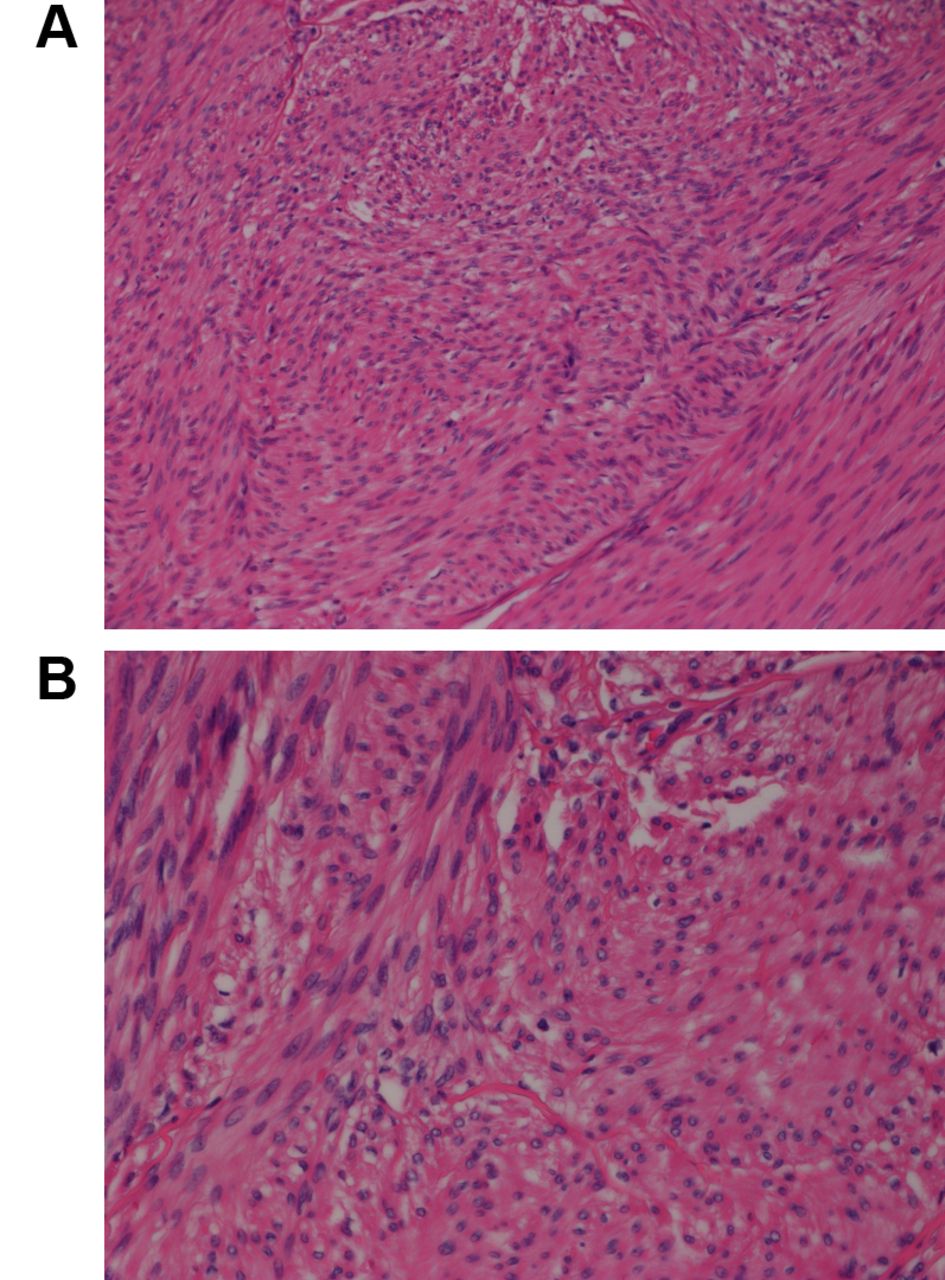
Microscopic examination of the spermatic cord leiomyoma. Hematoxylin and eosin-stained section showing bundles of elongated cells with cigar-shaped nuclei and eosinophilic cytoplasm. Original magnification, A: 20×; B: 40×.

G-Banding karyotyping and array comparative genomic hybridization (aCGH). analyses. Fresh tissue from a representative area of the tumor was analyzed cytogenetically as previously described (25). Genomic DNA was extracted from the tumor using the Maxwell RSC Instrument and Maxwell RSC Tissue DNA Kit (Promega, Madison, WI, USA), the concentration was measured using Quantus Fluorometer and the QuantiFluor ONE dsDNA System (Promega), and aCGH was performed using CytoSure array products (Oxford Gene Technology, Begbroke, Oxfordshire, UK) as previously described (26).

RNA sequencing. Total RNA was extracted from frozen tumor tissue adjacent to that used for cytogenetic analysis and histological examination using miRNeasy Mini Kit (Qiagen, Hilden, Germany). One microgram of total RNA from the tumor was sent to the Genomics Core Facility at the Norwegian Radium Hospital, Oslo University Hospital for high-throughput paired-end RNA-sequencing. Fusion transcripts were found using FusionCatcher software (27, 28).

Reverse transcription PCR (RT-PCR) and Sanger sequencing analyses. The primers used for PCR amplification and Sanger sequencing analyses are shown in Table I. The primer combinations and the corresponding fusion transcripts are presented in Table II. The iScript Advanced cDNA Synthesis Kit for RT-PCR was used to reverse transcribe 1 μg of total RNA according to the manufacturer’s instructions (Bio-Rad, Hercules, CA, USA). cDNA corresponding to 20 ng total RNA was used as template in subsequent PCR assays. BigDye Direct Cycle Sequencing Kit was used to perform both PCR and cycle (Sanger) sequencing according to the company’s recommendations (ThermoFisher Scientific, Waltham, MA, USA). Sequencing was run on an Applied Biosystems SeqStudio Genetic Analyzer system (ThermoFisher Scientific). For computer analysis of sequence data, the basic local alignment search tool (BLAST) software was used (29).

View this table:
  • View inline
  • View popup
  • Download powerpoint
Table I.

Primers used for polymerase chain reaction amplification and Sanger sequencing analyses. The forward primers (F1 and F2) had at their 5’-ends the M13 forward primer sequence: 5’-TGTAAAACGACGGCCAGT-3’. The reverse primers (R1) had at their 5’-ends the M13 reverse primer sequence: 5’-CAGGAAACAGCTATGACC-3’.

View this table:
  • View inline
  • View popup
  • Download powerpoint
Table II.

Primer combinations for polymerase chain reaction amplification to detect fusion transcripts.

Results

The G-banding analysis (Figure 2A) yielded the following karyotype: 48~50,XY,add(3)(p21),+4,+7,+8,+9,add(21)(q22)[cp9]/46,XY[2].

Figure 2.
  • Download figure
  • Open in new tab
  • Download powerpoint
Figure 2.

Cytogenetic and array comparative genomic hybridization (aCGH) analyses of the spermatic cord leiomyoma. A: Partial karyotype showing the add(3) and add(21) chromosomes with corresponding normal chromosome homologs. B: aCGH showing gains of one copy of each of chromosomes 4, 7, 8, and 9, as well as multiple gains and losses of genomic material from the p arm of chromosome 3 and q arm of chromosome 21. C: aCGH showing multiple gains and losses of genomic material from the p arm of chromosome 3. D: aCGH showing the multiple gain and losses on the q arm of chromosome 21.

aCGH confirmed trisomy for chromosomes 4,7,8, and 9 (Figure 2B). In addition, multiple gains and losses were detected on the p arm of chromosome 3 (Figure 2C) and on the q arm of chromosome 21 (Figure 2D).

RNA sequencing analysis with FusionCatcher detected seven fusion genes (Table III, Figure 3A). One transcript was detected for each of the chimeras inositol 1,4,5-trisphosphate receptor divergent transcript–nuclear receptor subfamily 2 group C member 2 (ITPR1–DT–NR2C2), cytoplasmic linker associated protein 2–interleukin 17 receptor D (CLASP2– IL17RD), nuclear receptor subfamily 2 group C member 2– cilia and flagella-associated protein 410 (NR2C2–CFAP410), zinc finger protein 621–leucyl-tRNA synthetase 2, mitochondrial (ZNF621–LARS2), and contactin 4–ras homolog family member A (CNTN4–RHOA), whereas two chimeric transcripts were detected for the fusion genes Rho guanine nucleotide exchange factor 3–calcium voltage-gated channel auxiliary subunit alpha2delta 2 (ARHGEF3– CACNA2D2) and trafficking kinesin protein 1–TIMP metallopeptidase inhibitor 4 (TRAK1–TIMP4) (Table III). In six of the fusion genes, both the 5’-end and 3’-end fusion partner genes were mapped on chromosome arm 3p (Table III, Figure 3A). In the seventh, the NR2C2-CFAP410 chimera, the 5’-end partner gene, NR2C2, was from 3p25.1, whereas CFAP410 was from 21q22.3 (Tables I and III). All fusion transcripts were verified using RT-PCR and Sanger sequencing analyses (Figure 3B).

View this table:
  • View inline
  • View popup
  • Download powerpoint
Table III.

Fusion transcripts detected in the spermatic cord leiomyoma using RNA sequencing and FusionCatcher.

Figure 3.
  • Download figure
  • Open in new tab
  • Download powerpoint
Figure 3.

The detected chimeras in the spermatic cord leiomyoma. A: Diagram of the p arm of chromosome 3, showing the mapping positions of the genes contactin 4 (CNTN4); inositol 1;4;5-trisphosphate receptor divergent transcript LITPR1-DT); TIMP metallopeptidase inhibitor 4 (TIMP4); nuclear receptor subfamily 2 group C member 2 (NR2C2); cytoplasmic linker associated protein 2 (CLASP2); zinc finger protein 621 (ZNF621); trafficking kinesin protein 1 (TRAK1); leucyl-tRNA synthetase 2, mitochondrial (LARS2); ras homolog family member A (RHOA); calcium voltage-gated channel auxiliary subunit alpha2delta 2 (CACNA2D2); Rho guanine nucleotide exchange factor 3 (ARHGEF3); and interleukin 17 receptor D (IL17RD); as well as the chimeric genes CNTN4−RHOA, ITPR1DT−NR2C2, TRAK1−TIMP4, CLASP2−IL17RD, ZNF621−LARS2 and ARHGEF3−CACNA2D2. The ↑ arrow next to a gene symbol indicates transcription of the gene from centromere to telomere. The ↓ arrow indicates transcription of the gene from telomere to centromere. B: Partial sequence chromatograms showing the junction positions in the chimeric transcripts CNTN4–RHOA, ITPR1-DT–NR2C2, NR2C2−CFAP410, CLASP2−IL17RD, ZNF621−LARS2, TRAK1−TIMP4 and ARHGEF3−CACNA2D2.

Discussion

Prior to the present study, to our knowledge, only a single para-testicular leiomyoma, located beneath the tunica albuginea, had been cytogenetically described, by Gorunova et al. (24). That tumor had an abnormal karyotype: 46,XY,der(5)t(5;14)(q31;q24),der(14)t(12;14)(q15; q24). The net imbalance of the structural rearrangements thus seemed to be the loss of 5q31qter and gain of 12q15qter. Molecular investigations were not performed, but strong nuclear immunostaining was found for the HMGA2 protein in the tumor cells, suggesting that the der(14)t(12;14)(q15;q24) dysregulated the expression of the HMGA2 gene. Because t(12;14)(q15;q24) is the most frequent translocation in uterine leiomyomas and leads to overexpression of HMGA2, the authors concluded that a common pathogenetic pathway exists for both para-testicular (male) and uterine (female) leiomyomas (24).

The present leiomyoma was found in the spermatic cord and had different cytogenetic aberrations from those described by Gorunova et al. (24). Trisomies were found for chromosomes 4, 7, 8, and 9, whereas structural aberrations affected chromosome arms 3p and 21q (Figure 2A). The observed numerical aberrations have also been reported in leiomyomas (30): trisomies for chromosomes 4, 8, and 9 in uterine leiomyomas (16, 31-35) and trisomy 7 in uterine, bladder, and gastric leiomyomas (16, 17, 31, 36).

Rearrangements of the 3p arm were reported in 22 uterine leiomyomas (30). In 12 of them, the 3p aberration was found together with rearrangements of chromosome bands 12q14~15 or band 6p21. Leiomyoma aberrations in the region 12q14~15 target the HMGA2 gene, whereas aberrations in 6p21 target HMGA1 (37, 38). Reports have been published on nine leiomyomas with aberration in the p arm of chromosome 3 without microscopically visible involvement of 12q14~15 or 6p21 (Table IV) (31, 32, 35, 39-42). Structural rearrangement of the 21q arm were reported in 12 uterine leiomyomas, always together with aberrations of 6p (often 6p21) or 12q14~15 (32, 38, 39, 43-46).

View this table:
  • View inline
  • View popup
  • Download powerpoint
Table IV.

Reported leiomyomas carrying a cytogenetic aberration in the p arm of chromosome 3.

The aCGH investigation of the tumor in our case not only confirmed trisomies for chromosomes 4,7, 8, and 9, but also showed multiple gains and losses of material from chromosome arms 3p and 21q (Figure 2B-D), suggesting that the rearrangements detected in 3p and 21q, add(3)(p21) and add(21)(q22) were more complex than those seen in G-banded preparations. RNA sequencing detected six fusion genes, ARHGEF3–CACNA2D2, TRAK1–TIMP4, ITPR1– DT–NR2C2, CLASP2–IL17RD, ZNF621–LARS2, and CNTN4–RHOA, with both 5’- and 3’-end fusion partner genes located on 3p (Table III and Figure 3A), and one fusion gene, NR2C2–CFAP410, with the 5’-end partner (NR2C2) from 3p and the 3’-end partner gene (CFAP410) from 21q (Table III). All of them were verified by RT-PCR/Sanger sequencing methodologies (Tables I and II; Figure 3B).

The data indicated that chromothripsis involving chromosome arms 3p and 21q occurred in the tumor, causing the generation of multiple fusion genes (47, 48). In this phenomenon, through some unusual, cataclysmic event, a chromosome breaks into many fragments, whereupon these fragments, or some of them, are randomly reassembled (47, 48). Some chromosome fragments are lost through this process while some fusion genes with oncogenic potential are generated at the junctions of the assembled fragments (47, 48). Chromothripsis has been detected in uterine leiomyomas which had neither mutations in MED12 or fumarate hydratase genes, nor 12q14~15 or 6p21 chromosomal rearrangements (49-52).

Three types of chimeras were found in our case of para-testicular leiomyoma: The first type, illustrated by the fusions CNTN4−RHOA, ITPR1-DT−NR2C2, and NR2C2−CFAP410, was the promoter-replacement chimera in which 5’-end untranslated regions of the 5’-end fusion partner gene replaced the 5’-end untranslated regions of the 3’-end fusion partner gene. Consequently, the 3’-end fusion partner comes under the control of the 5’-end fusion partner gene promoter. Thus, in the CNTN4–RHOA transcript, the first untranslated exon of CNTN4, which is highly expressed in testis, replaced the first untranslated exon of RHOA, bringing the entire coding part of RHOA under control of the CNTN4 promoter (53). RHOA codes for a member of the Rho family of small GTPases with multiple cellular and biological functions (54-58). Dysregulation of RHOA and other members of the Rho family of small GTPases has been reported in many neoplasias (59, 60).

In ITPR1-DT−NR2C2, the first untranslated exon of ITPR1-DT, which is part of the ITPR1 promoter, replaced the first untranslated exon of NR2C2 (61, 62). Consequently, the coding part of NR2C2 came under the control of the ITPR1 promoter. NR2C2 protein belongs to the nuclear hormone receptor family, acts as a ligand-activated transcription factor and plays a role in many biological processes such as development, cellular differentiation, and homeostasis (63). In the NR2C2–CFAP410 chimeric transcript, the first untranslated exon of NR2C2 fused to exon 4 of CFAP410. In exon 4 of CFAP410, there is an ATG which could act as a starting codon (NM_004928.3; position 521-523; XM_017028472.1; 421-423). In NR2C2-CFAP410, the part of CFAP410 coding for the last 147 amino acids (positions 109-256 in the sequence with accession number NP_0049 19.1; protein with accession number XP_016883961.1) comes under the control of the NR2C2 promoter. Four alternatively spliced transcript variants have been found for the CFAP410 gene. Three of the transcript variants code for mitochondrial proteins. The fourth transcript (accession number NM_001271442.1) codes for a cytoplasmic protein lacking the mitochondrion-target peptide (NP_001258371.1; https://www.ncbi.nlm.nih.gov/gene/755). CFAP410 was found to form a complex with NIMA-related serine/threonine kinase 1 (NEK1) protein and is involved in DNA-damage repair (64). Similarly to the fourth transcript variant of CFAP410, the putative CFAP410 translated from the NR2C2– CFAP410 chimera of this tumor would also be expected to lack the mitochondrion-target peptide and might interfere with the CFAP410/NEK1 complex and DNA-damage repair.

The second type of chimera involves out-of-frame transcripts. In the two detected ARHGEF3–CACNA2D2 transcripts, exon 1 or 2 of ARHGEF3 fused with exon 2 of CACNA2D2, introducing a stop codon shortly after the junctions. ARHGEF3 (also known as XPLN) codes for guanine nucleotide exchange factor 3, which specifically activates Rho GTPase family members RHOA and RHOB (65, 66). The putative truncated form of ARHGEF3 would lack the carboxyl terminal part of the ARHGEF3 protein responsible for guanine nucleotide exchange factor activity but retain the amino terminal part of the protein which interacts with mammalian target of rapamycin complex 2 (mTORC2), inhibiting mTORC2 kinase activity (66, 67). The presence of the truncated ARGEF3 protein and its possible functions cannot be determined without further cellular studies. Nevertheless, the ARHGEF3–CACNA2D2 transcript indicated further the dysfunction of RHOA signal transduction in this tumor.

The third type of chimera resulted in in-frame transcripts translated into chimeric proteins. In the ZNF621–LARS2 transcript, exon 2 of ZNF621 fused in-frame to exon 5 of LARS2. In the chimeric ZNF621–LARS2, the first 121 amino acids of LARS2 are replaced by the first eight amino acids of ZNF621. The LARS2 gene codes for a mitochondrial leucyl-tRNA synthetase with a mitochondrial targeting signal within its first 50 amino acids (68). Thus, since the mitochondrial targeting signal is absent from the chimeric ZNF621–LARS2 protein, the protein would probably be unable to enter the mitochondrion from the cytosol.

The two in-frame TRAK1–TIMP4 fusion transcripts would also give rise to chimeric proteins. The ubiquitously expressed TRAK1 gene codes for trafficking kinesin-binding protein 1 which is involved in mitochondrial and endosome-to-lysosome trafficking (69-74). Based on the reference sequence NP_001036111.1, the 953 amino acids-long TRAK1 protein has the following regions: A huntingtin-associated protein 1 conserved region between amino acids 50-353 binding to huntingtin in a polyglutamine repeat-length-dependent manner; a region between amino acids 359-509 which interacts with hepatocyte growth factor-regulated tyrosine kinase substrate protein; a region named milton between 416-583 which recruits the heavy chain of kinesin to mitochondria enabling the motor movement function of kinesin; and a region between amino acids 658-672 which interacts with O-linked N-acetylglucosamine (GlcNAc) transferase protein (70-72, 75, 76).

TIMP4 which codes for tissue inhibitor metalloproteinase 4, is weakly expressed in testis (77) and inhibits many metalloproteinases (78-80). The inhibitory property of TIMP4 was shown to depend on its N-terminal part, between residues 1-127, which contains a site of interaction with the catalytic domain of metalloproteinases (79, 80). In the tumor investigated here, the two translated TRAK1–TIMP4 chimeric proteins would lack all the above-mentioned functional regions of TRAK1 and residues 1-127 of TIMP4 which interact with and inhibit metalloproteinases. Their role in tumorigenesis is difficult to assess.

Based on the reference sequences NM_015097.2/NP_055912.2 for CLASP2 and NM_017563.4/NP_060033.3 for IL17RD, the CLASP2–IL17RD chimeric transcript would be expected to consist of exons 1 to 34 of CLASP2 and exons 2 to 13 of IL17RD, be 12317 bp-long, and code for an 1,892 amino acid protein composed of amino acids 1 to 1,195 from CLASP2 and 43 to 739 from IL17RD. The CLASP2 gene codes for cytoplasmic linker protein-associating protein that is a microtubule plus end tracking protein (81, 82). CLASP2 is localized to the distal ends of microtubules and is involved in the regulation of microtubule dynamics (83). Microtubules switch between phases of growth and shrinkage through transitions known as microtubule catastrophe and rescue (84). CLASP2 was found to specifically suppress microtubule catastrophe and promote rescue without affecting the rates of microtubule growth or shrinkage (85, 86). CLASP2 functions in various microtubule-dependent processes including cell division, cytoskeletal remodeling for cell migration, and vesicle transport between intracellular structures and the plasma membrane (87). CLASP2 protein has multiple HEAT repeats, a structural motif composed of two alpha helices linked by a short loop (88), two domains from tumour overexpressed gene that interact with αβ-tubulin, contributing to microtubule dynamics (89, 90), a region for interaction of microtubule-associated protein RP/EB family member 1 (MAPRE1, also known as EB1) (85, 91), and a carboxyl terminal region which interacts with CLIP1 protein and is required for the localization of CLASP2 protein to the Golgi apparatus and kinetochores (92).

The IL17RD gene codes for a single pass transmembrane protein mainly located in plasma membrane and regulating various signal pathways such as fibroblast growth factor and mitogen-activated protein kinase/extracellular signal-regulated kinase signal pathways (93-95). Murine models and expression patterns indicate that IL17RD is a tumor-suppressor gene (96-100). Thus, CLASP2–IL17RD chimeric transcript would produce a chimeric protein affecting microtubules and signaling pathways.

In summary, our data, together with those previously published, indicate the existence of a group of leiomyomas cytogenetically characterized by aberration of the p arm of chromosome 3 and the formation of fusion genes. A chromothripsis event seems to lie behind the pathogenetic changes that occurred in this para-testicular leiomyoma, resulting in multiple fusion genes.

Acknowledgements

This work was supported by grants from Radiumhospitalets Legater.

Footnotes

  • Authors’ Contributions

    IP designed and supervised the research, performed molecular genetic and bioinformatic analyses, and wrote the article. LG performed cytogenetic analyses. KA performed molecular genetic analyses and evaluated the data. IL performed the pathological examination. SH assisted with experimental design and writing of the article. All Authors read and approved of the final article.

  • This article is freely accessible online.

  • Conflicts of Interest

    No potential conflicts of interest exist.

  • Received April 18, 2021.
  • Revision received May 11, 2021.
  • Accepted May 18, 2021.
  • Copyright© 2021, International Institute of Anticancer Research (Dr. George J. Delinasios), All rights reserved

References

  1. ↵
    1. Zimmermann A,
    2. Bernuit D,
    3. Gerlinger C,
    4. Schaefers M and
    5. Geppert K
    : Prevalence, symptoms and management of uterine fibroids: an international internet-based survey of 21,746 women. BMC Womens Health 12: 6, 2012. PMID: 22448610. DOI: 10.1186/1472-6874-12-6
    OpenUrlCrossRefPubMed
    1. Giuliani E,
    2. As-Sanie S and
    3. Marsh EE
    : Epidemiology and management of uterine fibroids. Int J Gynaecol Obstet 149(1): 3-9, 2020. PMID: 31960950. DOI: 10.1002/ijgo.13102
    OpenUrlCrossRefPubMed
  2. ↵
    1. Billings SD,
    2. Folpe AL and
    3. Weiss SW
    : Do leiomyomas of deep soft tissue exist? An analysis of highly differentiated smooth muscle tumors of deep soft tissue supporting two distinct subtypes. Am J Surg Pathol 25(9): 1134-1142, 2001. PMID: 11688572. DOI: 10.1097/00000478-200109000-00003
    OpenUrlCrossRefPubMed
    1. Fasih N,
    2. Prasad Shanbhogue AK,
    3. Macdonald DB,
    4. Fraser-Hill MA,
    5. Papadatos D,
    6. Kielar AZ,
    7. Doherty GP,
    8. Walsh C,
    9. McInnes M and
    10. Atri M
    : Leiomyomas beyond the uterus: unusual locations, rare manifestations. Radiographics 28(7): 1931-1948, 2008. PMID: 19001649. DOI: 10.1148/rg.287085095
    OpenUrlCrossRefPubMed
  3. ↵
    1. Miettinen M
    : Smooth muscle tumors of soft tissue and non-uterine viscera: biology and prognosis. Mod Pathol 27(Suppl 1): S17-S29, 2014. PMID: 24384850. DOI: 10.1038/modpathol.2013.178
    OpenUrlCrossRefPubMed
  4. ↵
    1. Commandeur AE,
    2. Styer AK and
    3. Teixeira JM
    : Epidemiological and genetic clues for molecular mechanisms involved in uterine leiomyoma development and growth. Hum Reprod Update 21(5): 593-615, 2015. PMID: 26141720. DOI: 10.1093/humupd/dmv030
    OpenUrlCrossRefPubMed
  5. ↵
    1. Laganà AS,
    2. Vergara D,
    3. Favilli A,
    4. La Rosa VL,
    5. Tinelli A,
    6. Gerli S,
    7. Noventa M,
    8. Vitagliano A,
    9. Triolo O,
    10. Rapisarda AMC and
    11. Vitale SG
    : Epigenetic and genetic landscape of uterine leiomyomas: a current view over a common gynecological disease. Arch Gynecol Obstet 296(5): 855-867, 2017. PMID: 28875276. DOI: 10.1007/s00404-017-4515-5
    OpenUrlCrossRefPubMed
    1. McWilliams MM and
    2. Chennathukuzhi VM
    : Recent advances in uterine fibroid etiology. Semin Reprod Med 35(2): 181-189, 2017. PMID: 28278535. DOI: 10.1055/s-0037-1599090
    OpenUrlCrossRefPubMed
  6. ↵
    1. Baranov VS,
    2. Osinovskaya NS and
    3. Yarmolinskaya MI
    : Pathogenomics of uterine fibroids development. Int J Mol Sci 20(24): 6151, 2019. PMID: 31817606. DOI: 10.3390/ijms20246151
    OpenUrlCrossRefPubMed
  7. ↵
    1. Nibert M and
    2. Heim S
    : Uterine leiomyoma cytogenetics. Genes Chromosomes Cancer 2(1): 3-13, 1990. PMID: 2278965. DOI: 10.1002/gcc.2870020103
    OpenUrlCrossRefPubMed
  8. ↵
    1. Ligon AH and
    2. Morton CC
    : Leiomyomata: heritability and cytogenetic studies. Hum Reprod Update 7(1): 8-14, 2001. PMID: 11212080. DOI: 10.1093/humupd/7.1.8
    OpenUrlCrossRefPubMed
  9. ↵
    1. Mäkinen N,
    2. Heinonen HR,
    3. Moore S,
    4. Tomlinson IP,
    5. van der Spuy ZM and
    6. Aaltonen LA
    : MED12 exon 2 mutations are common in uterine leiomyomas from South African patients. Oncotarget 2(12): 966-969, 2011. PMID: 22182697. DOI: 10.18632/oncotarget.370
    OpenUrlCrossRefPubMed
    1. Mäkinen N,
    2. Mehine M,
    3. Tolvanen J,
    4. Kaasinen E,
    5. Li Y,
    6. Lehtonen HJ,
    7. Gentile M,
    8. Yan J,
    9. Enge M,
    10. Taipale M,
    11. Aavikko M,
    12. Katainen R,
    13. Virolainen E,
    14. Böhling T,
    15. Koski TA,
    16. Launonen V,
    17. Sjöberg J,
    18. Taipale J,
    19. Vahteristo P and
    20. Aaltonen LA
    : MED12, the mediator complex subunit 12 gene, is mutated at high frequency in uterine leiomyomas. Science 334(6053): 252-255, 2011. PMID: 21868628. DOI: 10.1126/science.1208930
    OpenUrlAbstract/FREE Full Text
  10. ↵
    1. Heinonen HR,
    2. Sarvilinna NS,
    3. Sjöberg J,
    4. Kämpjärvi K,
    5. Pitkänen E,
    6. Vahteristo P,
    7. Mäkinen N and
    8. Aaltonen LA
    : MED12 mutation frequency in unselected sporadic uterine leiomyomas. Fertil Steril 102(4): 1137-1142, 2014. PMID: 25108465. DOI: 10.1016/j.fertnstert.2014.06.040
    OpenUrlCrossRefPubMed
  11. ↵
    1. Mehine M,
    2. Kaasinen E,
    3. Heinonen HR,
    4. Mäkinen N,
    5. Kämpjärvi K,
    6. Sarvilinna N,
    7. Aavikko M,
    8. Vähärautio A,
    9. Pasanen A,
    10. Bützow R,
    11. Heikinheimo O,
    12. Sjöberg J,
    13. Pitkänen E,
    14. Vahteristo P and
    15. Aaltonen LA
    : Integrated data analysis reveals uterine leiomyoma subtypes with distinct driver pathways and biomarkers. Proc Natl Acad Sci USA 113(5): 1315-1320, 2016. PMID: 26787895. DOI: 10.1073/pnas.1518752113
    OpenUrlAbstract/FREE Full Text
  12. ↵
    1. Teyssier JR and
    2. Ferre D
    : Frequent clonal chromosomal changes in human non-malignant tumors. Int J Cancer 44(5): 828-832, 1989. PMID: 2583864. DOI: 10.1002/ijc.2910440514
    OpenUrlCrossRefPubMed
  13. ↵
    1. Bardi G,
    2. Johansson B,
    3. Pandis N,
    4. Heim S,
    5. Mandahl N,
    6. Bak-Jensen E,
    7. Frederiksen H,
    8. Andrén-Sandberg A and
    9. Mitelman F
    : Recurrent chromosome aberrations in abdominal smooth muscle tumors. Cancer Genet Cytogenet 62(1): 43-46, 1992. PMID: 1521232. DOI: 10.1016/0165-4608(92)90036-8
    OpenUrlCrossRefPubMed
    1. Dal Cin P,
    2. Aly MS,
    3. Dewever I,
    4. Vandamme B and
    5. Vandenberghe H
    : Does chromosome investigation discriminate between benign and malignant gastrointestinal leiomyomatous tumors? Diag Oncol 2(1): 55-59, 1992.
    OpenUrl
    1. Heidet L,
    2. Boye E,
    3. Cai Y,
    4. Sado Y,
    5. Zhang X,
    6. Fléjou JF,
    7. Fékété F,
    8. Ninomiya Y,
    9. Gubler MC and
    10. Antignac C
    : Somatic deletion of the 5’ ends of both the COL4A5 and COL4A6 genes in a sporadic leiomyoma of the esophagus. Am J Pathol 152(3): 673-678, 1998. PMID: 9502408.
    OpenUrlPubMed
  14. ↵
    1. Panagopoulos I,
    2. Gorunova L,
    3. Lund-Iversen M,
    4. Lobmaier I,
    5. Bjerkehagen B and
    6. Heim S
    : Recurrent fusion of the genes FN1 and ALK in gastrointestinal leiomyomas. Mod Pathol 29(11): 1415-1423, 2016. PMID: 27469327. DOI: 10.1038/modpathol.2016.129
    OpenUrlCrossRefPubMed
  15. ↵
    1. Panagopoulos I,
    2. Gorunova L,
    3. Bjerkehagen B and
    4. Heim S
    : Fusion of the genes EWSR1 and PBX3 in retroperitoneal leiomyoma with t(9;22)(q33;q12). PLoS One 10(4): e0124288, 2015. PMID: 25875009. DOI: 10.1371/journal.pone.0124288
    OpenUrlCrossRefPubMed
    1. Panagopoulos I,
    2. Gorunova L,
    3. Bjerkehagen B and
    4. Heim S
    : Novel KAT6B-KANSL1 fusion gene identified by RNA sequencing in retroperitoneal leiomyoma with t(10;17)(q22;q21). PLoS One 10(1): e0117010, 2015. PMID: 25621995. DOI: 10.1371/journal.pone.0117010
    OpenUrlCrossRefPubMed
  16. ↵
    1. Panagopoulos I,
    2. Gorunova L,
    3. Brunetti M,
    4. Agostini A,
    5. Andersen HK,
    6. Lobmaier I,
    7. Bjerkehagen B and
    8. Heim S
    : Genetic heterogeneity in leiomyomas of deep soft tissue. Oncotarget 8(30): 48769-48781, 2017. PMID: 28591699. DOI: 10.18632/oncotarget.17953
    OpenUrlCrossRefPubMed
  17. ↵
    1. Gorunova L,
    2. Bjerkehagen B and
    3. Heim S
    : Paratesticular leiomyoma with a der(14)t(12;14)(q15;q24). Cancer Genet 204(8): 465-468, 2011. PMID: 21962898. DOI: 10.1016/j.cancergen.2011.06.005
    OpenUrlCrossRefPubMed
  18. ↵
    1. Panagopoulos I,
    2. Gorunova L,
    3. Lund-Iversen M,
    4. Andersen K,
    5. Andersen HK,
    6. Lobmaier I,
    7. Bjerkehagen B and
    8. Heim S
    : Cytogenetics of spindle cell/pleomorphic lipomas: Karyotyping and FISH analysis of 31 tumors. Cancer Genomics Proteomics 15(3): 193-200, 2018. PMID: 29695401. DOI: 10.21873/cgp.20077
    OpenUrlAbstract/FREE Full Text
  19. ↵
    1. Panagopoulos I,
    2. Gorunova L,
    3. Andersen K,
    4. Lund-Iversen M,
    5. Lobmaier I,
    6. Micci F and
    7. Heim S
    : NDRG1-PLAG1 and TRPS1-PLAG1 fusion genes in chondroid syringoma. Cancer Genomics Proteomics 17(3): 237-248, 2020. PMID: 32345665. DOI: 10.21873/cgp.20184
    OpenUrlAbstract/FREE Full Text
  20. ↵
    1. Kangaspeska S,
    2. Hultsch S,
    3. Edgren H,
    4. Nicorici D,
    5. Murumägi A and
    6. Kallioniemi O
    : Reanalysis of RNA-sequencing data reveals several additional fusion genes with multiple isoforms. PLoS One 7(10): e48745, 2012. PMID: 23119097. DOI: 10.1371/journal.pone.0048745
    OpenUrlCrossRefPubMed
  21. ↵
    1. Nicorici D,
    2. Satalan M,
    3. Edgren H,
    4. Kangaspeska S,
    5. Murumagi A,
    6. Kallioniemi O,
    7. Virtanen S and
    8. Kilkku O
    : FusionCatcher – a tool for finding somatic fusion genes in paired-end RNA-sequencing data. bioRxiv, 2014. DOI: 10.1101/011650
    OpenUrlAbstract/FREE Full Text
  22. ↵
    1. Altschul SF,
    2. Gish W,
    3. Miller W,
    4. Myers EW and
    5. Lipman DJ
    : Basic local alignment search tool. J Mol Biol 215(3): 403-410, 1990. PMID: 2231712. DOI: 10.1016/S0022-2836(05)80360-2
    OpenUrlCrossRefPubMed
  23. ↵
    1. Mitelman F,
    2. Johansson B and
    3. Mertens F
    : Mitelman Database of Chromosome Aberrations and Gene Fusions in Cancer, 2021. Available at: https://mitelmandatabase.isb-cgc.org/ [Last accessed on April 25, 2021]
  24. ↵
    1. Hu J and
    2. Surti U
    : Subgroups of uterine leiomyomas based on cytogenetic analysis. Hum Pathol 22(10): 1009-1016, 1991. PMID: 1842373. DOI: 10.1016/0046-8177(91)90009-e
    OpenUrlCrossRefPubMed
  25. ↵
    1. Nilbert M,
    2. Heim S,
    3. Mandahl N,
    4. Flodérus UM,
    5. Willén H and
    6. Mitelman F
    : Characteristic chromosome abnormalities, including rearrangements of 6p, del(7q), +12, and t(12;14), in 44 uterine leiomyomas. Hum Genet 85(6): 605-611, 1990. PMID: 2227952. DOI: 10.1007/BF00193583
    OpenUrlCrossRefPubMed
    1. Nilbert M,
    2. Heim S,
    3. Mandahl N,
    4. Flodérus UM,
    5. Willén H and
    6. Mitelman F
    : Trisomy 12 in uterine leiomyomas. A new cytogenetic subgroup. Cancer Genet Cytogenet 45(1): 63-66, 1990. PMID: 2302686. DOI: 10.1016/0165-4608(90)90067-k
    OpenUrlCrossRefPubMed
    1. Christacos NC,
    2. Quade BJ,
    3. Dal Cin P and
    4. Morton CC
    : Uterine leiomyomata with deletions of Ip represent a distinct cytogenetic subgroup associated with unusual histologic features. Genes Chromosomes Cancer 45(3): 304-312, 2006. PMID: 16320247. DOI: 10.1002/gcc.20291
    OpenUrlCrossRefPubMed
  26. ↵
    1. Meloni AM,
    2. Surti U,
    3. Contento AM,
    4. Davare J and
    5. Sandberg AA
    : Uterine leiomyomas: cytogenetic and histologic profile. Obstet Gynecol 80(2): 209-217, 1992. PMID: 1635734.
    OpenUrlPubMed
  27. ↵
    1. Horton ES,
    2. Dobin SM and
    3. Donner LR
    : Leiomyoma of the urinary bladder: a cytogenetic study of a case. Cancer Genet Cytogenet 177(2): 147-148, 2007. PMID: 17854672. DOI: 10.1016/j.cancergencyto.2007.06.009
    OpenUrlCrossRefPubMed
  28. ↵
    1. Klemke M,
    2. Meyer A,
    3. Nezhad MH,
    4. Bartnitzke S,
    5. Drieschner N,
    6. Frantzen C,
    7. Schmidt EH,
    8. Belge G and
    9. Bullerdiek J
    : Overexpression of HMGA2 in uterine leiomyomas points to its general role for the pathogenesis of the disease. Genes Chromosomes Cancer 48(2): 171-178, 2009. PMID: 18980243. DOI: 10.1002/gcc.20627
    OpenUrlCrossRefPubMed
  29. ↵
    1. Nezhad MH,
    2. Drieschner N,
    3. Helms S,
    4. Meyer A,
    5. Tadayyon M,
    6. Klemke M,
    7. Belge G,
    8. Bartnitzke S,
    9. Burchardt K,
    10. Frantzen C,
    11. Schmidt EH and
    12. Bullerdiek J
    : 6p21 rearrangements in uterine leiomyomas targeting HMGA1. Cancer Genet Cytogenet 203(2): 247-252, 2010. PMID: 21156240. DOI: 10.1016/j.cancergencyto.2010.08.005
    OpenUrlCrossRefPubMed
  30. ↵
    1. Karaiskos C,
    2. Pandis N,
    3. Bardi G,
    4. Sfikas K,
    5. Tserkezoglou A,
    6. Fotiou S and
    7. Heim S
    : Cytogenetic findings in uterine epithelioid leiomyomas. Cancer Genet Cytogenet 80(2): 103-106, 1995. PMID: 7736423. DOI: 10.1016/0165-4608(94)00167-a
    OpenUrlCrossRefPubMed
    1. Nilbert M,
    2. Heim S,
    3. Mandahl N,
    4. Flodérus UM,
    5. Willén H and
    6. Mitelman F
    : Karyotypic rearrangements in 20 uterine leiomyomas. Cytogenet Cell Genet 49(4): 300-304, 1988. PMID: 3248388. DOI: 10.1159/000132682
    OpenUrlCrossRefPubMed
    1. Stern C,
    2. Deichert U,
    3. Thode B,
    4. Bartnitzke S and
    5. Bullerdiek J
    : Cytogenetic subtyping of 139 uterine leiomyoma. Geburtshilfe Frauenheilkd 52(12): 767-772, 1992. PMID: 1490556. DOI: 10.1055/s-2007-1023809
    OpenUrlCrossRefPubMed
  31. ↵
    1. Vanni R,
    2. Lecca U and
    3. Faa G
    : Uterine leiomyoma cytogenetics. II. Report of forty cases. Cancer Genet Cytogenet 53(2): 247-256, 1991. PMID: 2065298. DOI: 10.1016/0165-4608(91)90101-y
    OpenUrlCrossRefPubMed
  32. ↵
    1. Mark J,
    2. Havel G,
    3. Dahlenfors R and
    4. Wedell B
    : Cytogenetics of multiple uterine leiomyomas, parametrial leiomyoma and disseminated peritoneal leiomyomatosis. Anticancer Res 11(1): 33-39, 1991. PMID: 2018368.
    OpenUrlPubMed
    1. Mark J,
    2. Havel G,
    3. Grepp C,
    4. Dahlenfors R and
    5. Wedell B
    : Chromosomal patterns in human benign uterine leiomyomas. Cancer Genet Cytogenet 44(1): 1-13, 1990. PMID: 2293875. DOI: 10.1016/0165-4608(90)90192-d
    OpenUrlCrossRefPubMed
    1. Pandis N,
    2. Heim S,
    3. Bardi G,
    4. Flodérus UM,
    5. Willén H,
    6. Mandahl N and
    7. Mitelman F
    : Chromosome analysis of 96 uterine leiomyomas. Cancer Genet Cytogenet 55(1): 11-18, 1991. PMID: 1913597. DOI: 10.1016/0165-4608(91)90229-n
    OpenUrlCrossRefPubMed
  33. ↵
    1. Rein MS,
    2. Powell WL,
    3. Walters FC,
    4. Weremowicz S,
    5. Cantor RM,
    6. Barbieri RL and
    7. Morton CC
    : Cytogenetic abnormalities in uterine myomas are associated with myoma size. Mol Hum Reprod 4(1): 83-86, 1998. PMID: 9510016. DOI: 10.1093/molehr/4.1.83
    OpenUrlCrossRefPubMed
  34. ↵
    1. Korbel JO and
    2. Campbell PJ
    : Criteria for inference of chromothripsis in cancer genomes. Cell 152(6): 1226-1236, 2013. PMID: 23498933. DOI: 10.1016/j.cell.2013.02.023
    OpenUrlCrossRefPubMed
  35. ↵
    1. Koltsova AS,
    2. Pendina AA,
    3. Efimova OA,
    4. Chiryaeva OG,
    5. Kuznetzova TV and
    6. Baranov VS
    : On the complexity of mechanisms and consequences of chromothripsis: an update. Front Genet 10: 393, 2019. PMID: 31114609. DOI: 10.3389/fgene.2019.00393
    OpenUrlCrossRefPubMed
  36. ↵
    1. Mehine M,
    2. Kaasinen E,
    3. Mäkinen N,
    4. Katainen R,
    5. Kämpjärvi K,
    6. Pitkänen E,
    7. Heinonen HR,
    8. Bützow R,
    9. Kilpivaara O,
    10. Kuosmanen A,
    11. Ristolainen H,
    12. Gentile M,
    13. Sjöberg J,
    14. Vahteristo P and
    15. Aaltonen LA
    : Characterization of uterine leiomyomas by whole-genome sequencing. N Engl J Med 369(1): 43-53, 2013. PMID: 23738515. DOI: 10.1056/NEJMoa1302736
    OpenUrlCrossRefPubMed
    1. Holzmann C,
    2. Markowski DN,
    3. Koczan D,
    4. Küpker W,
    5. Helmke BM and
    6. Bullerdiek J
    : Cytogenetically normal uterine leiomyomas without MED12-mutations - a source to identify unknown mechanisms of the development of uterine smooth muscle tumors. Mol Cytogenet 7(1): 88, 2014. PMID: 25506394. DOI: 10.1186/s13039-014-0088-1
    OpenUrlCrossRefPubMed
    1. Mehine M,
    2. Mäkinen N,
    3. Heinonen HR,
    4. Aaltonen LA and
    5. Vahteristo P
    : Genomics of uterine leiomyomas: insights from high-throughput sequencing. Fertil Steril 102(3): 621-629, 2014. PMID: 25106763. DOI: 10.1016/j.fertnstert.2014.06.050
    OpenUrlCrossRefPubMed
  37. ↵
    1. Pendina AA,
    2. Koltsova AS,
    3. Efimova OA,
    4. Malysheva OV,
    5. Osinovskaya NS,
    6. Sultanov IY,
    7. Tikhonov AV,
    8. Shved NY,
    9. Chiryaeva OG,
    10. Simareva AD,
    11. Kakhiani MI and
    12. Baranov VS
    : Case of chromothripsis in a large solitary non-recurrent uterine leiomyoma. Eur J Obstet Gynecol Reprod Biol 219: 134-136, 2017. PMID: 29103617. DOI: 10.1016/j.ejogrb.2017.10.028
    OpenUrlCrossRefPubMed
  38. ↵
    1. Hansford LM,
    2. Smith SA,
    3. Haber M,
    4. Norris MD,
    5. Cheung B and
    6. Marshall GM
    : Cloning and characterization of the human neural cell adhesion molecule, CNTN4 (alias BIG-2). Cytogenet Genome Res 101(1): 17-23, 2003. PMID: 14571131. DOI: 10.1159/000073412
    OpenUrlCrossRefPubMed
  39. ↵
    1. Deng Z,
    2. Jia Y,
    3. Liu H,
    4. He M,
    5. Yang Y,
    6. Xiao W and
    7. Li Y
    : RhoA/ROCK pathway: implication in osteoarthritis and therapeutic targets. Am J Transl Res 11(9): 5324-5331, 2019. PMID: 31632513.
    OpenUrlPubMed
    1. Ueyama T
    : Rho-family small GTPases: From highly polarized sensory neurons to cancer cells. Cells 8(2): 92, 2019. PMID: 30696065. DOI: 10.3390/cells8020092
    OpenUrlCrossRefPubMed
    1. Kloc M,
    2. Uosef A,
    3. Kubiak JZ and
    4. Ghobrial RM
    : Role of macrophages and RhoA pathway in atherosclerosis. Int J Mol Sci 22(1): 216, 2020. PMID: 33379334. DOI: 10.3390/ijms22010216
    OpenUrlCrossRefPubMed
    1. Seccia TM,
    2. Rigato M,
    3. Ravarotto V and
    4. Calò LA
    : ROCK (RhoA/Rho Kinase) in cardiovascular-renal pathophysiology: A review of new advancements. J Clin Med 9(5): 1328, 2020. PMID: 32370294. DOI: 10.3390/jcm9051328
    OpenUrlCrossRefPubMed
  40. ↵
    1. Zhang Y,
    2. Saradna A,
    3. Ratan R,
    4. Ke X,
    5. Tu W,
    6. Do DC,
    7. Hu C and
    8. Gao P
    : RhoA/Rho-kinases in asthma: from pathogenesis to therapeutic targets. Clin Transl Immunology 9(5): e01134, 2020. PMID: 32355562. DOI: 10.1002/cti2.1134
    OpenUrlCrossRefPubMed
  41. ↵
    1. Aspenström P
    : Activated Rho GTPases in cancer-The beginning of a new paradigm. Int J Mol Sci 19(12): 3949, 2018. PMID: 30544828. DOI: 10.3390/ijms19123949
    OpenUrlCrossRefPubMed
  42. ↵
    1. Jung H,
    2. Yoon SR,
    3. Lim J,
    4. Cho HJ and
    5. Lee HG
    : Dysregulation of Rho GTPases in human cancers. Cancers (Basel) 12(5): 1179, 2020. PMID: 32392742. DOI: 10.3390/cancers12051179
    OpenUrlCrossRefPubMed
  43. ↵
    1. Kirkwood KL,
    2. Homick K,
    3. Dragon MB and
    4. Bradford PG
    : Cloning and characterization of the type I inositol 1,4,5-trisphosphate receptor gene promoter. Regulation by 17beta-estradiol in osteoblasts. J Biol Chem 272(36): 22425-22431, 1997. PMID: 9278393. DOI: 10.1074/jbc.272.36.22425
    OpenUrlAbstract/FREE Full Text
  44. ↵
    1. Deelman LE,
    2. Jonk LJ and
    3. Henning RH
    : The isolation and characterization of the promoter of the human type 1 inositol 1,4,5-trisphosphate receptor. Gene 207(2): 219-225, 1998. PMID: 9511764. DOI: 10.1016/s0378-1119(97)00630-6
    OpenUrlCrossRefPubMed
  45. ↵
    1. Lin SJ,
    2. Yang DR,
    3. Yang G,
    4. Lin CY,
    5. Chang HC,
    6. Li G and
    7. Chang C
    : TR2 and TR4 orphan nuclear receptors: an overview. Curr Top Dev Biol 125: 357-373, 2017. PMID: 28527578. DOI: 10.1016/bs.ctdb.2017.02.002
    OpenUrlCrossRefPubMed
  46. ↵
    1. Fang X,
    2. Lin H,
    3. Wang X,
    4. Zuo Q,
    5. Qin J and
    6. Zhang P
    : The NEK1 interactor, C21ORF2, is required for efficient DNA damage repair. Acta Biochim Biophys Sin (Shanghai) 47(10): 834-841, 2015. PMID: 26290490. DOI: 10.1093/abbs/gmv076
    OpenUrlCrossRefPubMed
  47. ↵
    1. Thiesen S,
    2. Kübart S,
    3. Ropers HH and
    4. Nothwang HG
    : Isolation of two novel human RhoGEFs, ARHGEF3 and ARHGEF4, in 3p13-21 and 2q22. Biochem Biophys Res Commun 273(1): 364-369, 2000. PMID: 10873612. DOI: 10.1006/bbrc.2000.2925
    OpenUrlCrossRefPubMed
  48. ↵
    1. Arthur WT,
    2. Ellerbroek SM,
    3. Der CJ,
    4. Burridge K and
    5. Wennerberg K
    : XPLN, a guanine nucleotide exchange factor for RhoA and RhoB, but not RhoC. J Biol Chem 277(45): 42964-42972, 2002. PMID: 12221096. DOI: 10.1074/jbc.M207401200
    OpenUrlAbstract/FREE Full Text
  49. ↵
    1. Khanna N,
    2. Fang Y,
    3. Yoon MS and
    4. Chen J
    : XPLN is an endogenous inhibitor of mTORC2. Proc Natl Acad Sci USA 110(40): 15979-15984, 2013. PMID: 24043828. DOI: 10.1073/pnas.1310434110
    OpenUrlAbstract/FREE Full Text
  50. ↵
    1. Bonnefond L,
    2. Fender A,
    3. Rudinger-Thirion J,
    4. Giegé R,
    5. Florentz C and
    6. Sissler M
    : Toward the full set of human mitochondrial aminoacyl-tRNA synthetases: characterization of AspRS and TyrRS. Biochemistry 44(12): 4805-4816, 2005. PMID: 15779907. DOI: 10.1021/bi047527z
    OpenUrlCrossRefPubMed
  51. ↵
    1. Stowers RS,
    2. Megeath LJ,
    3. Górska-Andrzejak J,
    4. Meinertzhagen IA and
    5. Schwarz TL
    : Axonal transport of mitochondria to synapses depends on milton, a novel Drosophila protein. Neuron 36(6): 1063-1077, 2002. PMID: 12495622. DOI: 10.1016/s0896-6273(02)01094-2
    OpenUrlCrossRefPubMed
  52. ↵
    1. Webber E,
    2. Li L and
    3. Chin LS
    : Hypertonia-associated protein Trak1 is a novel regulator of endosome-to-lysosome trafficking. J Mol Biol 382(3): 638-651, 2008. PMID: 18675823. DOI: 10.1016/j.jmb.2008.07.045
    OpenUrlCrossRefPubMed
    1. Brickley K,
    2. Pozo K and
    3. Stephenson FA
    : N-acetylglucosamine transferase is an integral component of a kinesin-directed mitochondrial trafficking complex. Biochim Biophys Acta 1813(1): 269-281, 2011. PMID: 21034780. DOI: 10.1016/j.bbamcr.2010.10.011
    OpenUrlCrossRefPubMed
  53. ↵
    1. Brickley K and
    2. Stephenson FA
    : Trafficking kinesin protein (TRAK)-mediated transport of mitochondria in axons of hippocampal neurons. J Biol Chem 286(20): 18079-18092, 2011. PMID: 21454691. DOI: 10.1074/jbc.M111.236018
    OpenUrlAbstract/FREE Full Text
    1. van Spronsen M,
    2. Mikhaylova M,
    3. Lipka J,
    4. Schlager MA,
    5. van den Heuvel DJ,
    6. Kuijpers M,
    7. Wulf PS,
    8. Keijzer N,
    9. Demmers J,
    10. Kapitein LC,
    11. Jaarsma D,
    12. Gerritsen HC,
    13. Akhmanova A and
    14. Hoogenraad CC
    : TRAK/Milton motor-adaptor proteins steer mitochondrial trafficking to axons and dendrites. Neuron 77(3): 485-502, 2013. PMID: 23395375. DOI: 10.1016/j.neuron.2012.11.027
    OpenUrlCrossRefPubMed
  54. ↵
    1. Barel O,
    2. Malicdan MCV,
    3. Ben-Zeev B,
    4. Kandel J,
    5. Pri-Chen H,
    6. Stephen J,
    7. Castro IG,
    8. Metz J,
    9. Atawa O,
    10. Moshkovitz S,
    11. Ganelin E,
    12. Barshack I,
    13. Polak-Charcon S,
    14. Nass D,
    15. Marek-Yagel D,
    16. Amariglio N,
    17. Shalva N,
    18. Vilboux T,
    19. Ferreira C,
    20. Pode-Shakked B,
    21. Heimer G,
    22. Hoffmann C,
    23. Yardeni T,
    24. Nissenkorn A,
    25. Avivi C,
    26. Eyal E,
    27. Kol N,
    28. Glick Saar E,
    29. Wallace DC,
    30. Gahl WA,
    31. Rechavi G,
    32. Schrader M,
    33. Eckmann DM and
    34. Anikster Y
    : Deleterious variants in TRAK1 disrupt mitochondrial movement and cause fatal encephalopathy. Brain 140(3): 568-581, 2017. PMID: 28364549. DOI: 10.1093/brain/awx002
    OpenUrlCrossRefPubMed
  55. ↵
    1. Iyer SP,
    2. Akimoto Y and
    3. Hart GW
    : Identification and cloning of a novel family of coiled-coil domain proteins that interact with O-GlcNAc transferase. J Biol Chem 278(7): 5399-5409, 2003. PMID: 12435728. DOI: 10.1074/jbc.M209384200
    OpenUrlAbstract/FREE Full Text
  56. ↵
    1. Pekkurnaz G,
    2. Trinidad JC,
    3. Wang X,
    4. Kong D and
    5. Schwarz TL
    : Glucose regulates mitochondrial motility via Milton modification by O-GlcNAc transferase. Cell 158(1): 54-68, 2014. PMID: 24995978. DOI: 10.1016/j.cell.2014.06.007
    OpenUrlCrossRefPubMed
  57. ↵
    1. Greene J,
    2. Wang M,
    3. Liu YE,
    4. Raymond LA,
    5. Rosen C and
    6. Shi YE
    : Molecular cloning and characterization of human tissue inhibitor of metalloproteinase 4. J Biol Chem 271(48): 30375-30380, 1996. PMID: 8939999. DOI: 10.1074/jbc.271.48.30375
    OpenUrlAbstract/FREE Full Text
  58. ↵
    1. Liu YE,
    2. Wang M,
    3. Greene J,
    4. Su J,
    5. Ullrich S,
    6. Li H,
    7. Sheng S,
    8. Alexander P,
    9. Sang QA and
    10. Shi YE
    : Preparation and characterization of recombinant tissue inhibitor of metalloproteinase 4 (TIMP-4). J Biol Chem 272(33): 20479-20483, 1997. PMID: 9252358. DOI: 10.1074/jbc.272.33.20479
    OpenUrlAbstract/FREE Full Text
  59. ↵
    1. Stratmann B,
    2. Farr M and
    3. Tschesche H
    : MMP-TIMP interaction depends on residue 2 in TIMP-4. FEBS Lett 507(3): 285-287, 2001. PMID: 11696356. DOI: 10.1016/s0014-5793(01)02987-8
    OpenUrlCrossRefPubMed
  60. ↵
    1. Stratmann B,
    2. Farr M and
    3. Tschesche H
    : Characterization of C-terminally truncated human tissue inhibitor of metalloproteinases-4 expressed in Pichia pastoris. Biol Chem 382(6): 987-991, 2001. PMID: 11501766. DOI: 10.1515/BC.2001.124
    OpenUrlCrossRefPubMed
  61. ↵
    1. Galjart N
    : Plus-end-tracking proteins and their interactions at microtubule ends. Curr Biol 20(12): R528-R537, 2010. PMID: 20620909. DOI: 10.1016/j.cub.2010.05.022
    OpenUrlCrossRefPubMed
  62. ↵
    1. Ferreira JG,
    2. Pereira AL and
    3. Maiato H
    : Microtubule plus-end tracking proteins and their roles in cell division. Int Rev Cell Mol Biol 309: 59-140, 2014. PMID: 24529722. DOI: 10.1016/B978-0-12-800255-1.00002-8
    OpenUrlCrossRefPubMed
  63. ↵
    1. Langlais P,
    2. Dillon JL,
    3. Mengos A,
    4. Baluch DP,
    5. Ardebili R,
    6. Miranda DN,
    7. Xie X,
    8. Heckmann BL,
    9. Liu J and
    10. Mandarino LJ
    : Identification of a role for CLASP2 in insulin action. J Biol Chem 287(46): 39245-39253, 2012. PMID: 22992739. DOI: 10.1074/jbc.M112.394148
    OpenUrlAbstract/FREE Full Text
  64. ↵
    1. Gardner MK,
    2. Zanic M and
    3. Howard J
    : Microtubule catastrophe and rescue. Curr Opin Cell Biol 25(1): 14-22, 2013. PMID: 23092753. DOI: 10.1016/j.ceb.2012.09.006
    OpenUrlCrossRefPubMed
  65. ↵
    1. Lawrence EJ,
    2. Arpag G,
    3. Norris SR and
    4. Zanic M
    : Human CLASP2 specifically regulates microtubule catastrophe and rescue. Mol Biol Cell 29(10): 1168-1177, 2018. PMID: 29540526. DOI: 10.1091/mbc.E18-01-0016
    OpenUrlCrossRefPubMed
  66. ↵
    1. Lawrence EJ and
    2. Zanic M
    : Rescuing microtubules from the brink of catastrophe: CLASPs lead the way. Curr Opin Cell Biol 56: 94-101, 2019. PMID: 30453184. DOI: 10.1016/j.ceb.2018.10.011
    OpenUrlCrossRefPubMed
  67. ↵
    1. Lawrence EJ,
    2. Zanic M and
    3. Rice LM
    : CLASPs at a glance. J Cell Sci 133(8): jcs243097, 2020. PMID: 32332092. DOI: 10.1242/jcs.243097
    OpenUrlAbstract/FREE Full Text
  68. ↵
    1. Yoshimura SH and
    2. Hirano T
    : HEAT repeats - versatile arrays of amphiphilic helices working in crowded environments? J Cell Sci 129(21): 3963-3970, 2016. PMID: 27802131. DOI: 10.1242/jcs.185710
    OpenUrlAbstract/FREE Full Text
  69. ↵
    1. Slep KC
    : The role of TOG domains in microtubule plus end dynamics. Biochem Soc Trans 37(Pt 5): 1002-1006, 2009. PMID: 19754440. DOI: 10.1042/BST0371002
    OpenUrlAbstract/FREE Full Text
  70. ↵
    1. Maki T,
    2. Grimaldi AD,
    3. Fuchigami S,
    4. Kaverina I and
    5. Hayashi I
    : CLASP2 has two distinct TOG domains that contribute differently to microtubule dynamics. J Mol Biol 427(14): 2379-2395, 2015. PMID: 26003921. DOI: 10.1016/j.jmb.2015.05.012
    OpenUrlCrossRefPubMed
  71. ↵
    1. Mimori-Kiyosue Y,
    2. Grigoriev I,
    3. Lansbergen G,
    4. Sasaki H,
    5. Matsui C,
    6. Severin F,
    7. Galjart N,
    8. Grosveld F,
    9. Vorobjev I,
    10. Tsukita S and
    11. Akhmanova A
    : CLASP1 and CLASP2 bind to EB1 and regulate microtubule plus-end dynamics at the cell cortex. J Cell Biol 168(1): 141-153, 2005. PMID: 15631994. DOI: 10.1083/jcb.200405094
    OpenUrlAbstract/FREE Full Text
  72. ↵
    1. Girão H,
    2. Okada N,
    3. Rodrigues TA,
    4. Silva AO,
    5. Figueiredo AC,
    6. Garcia Z,
    7. Moutinho-Santos T,
    8. Hayashi I,
    9. Azevedo JE,
    10. Macedo-Ribeiro S and
    11. Maiato H
    : CLASP2 binding to curved microtubule tips promotes flux and stabilizes kinetochore attachments. J Cell Biol 219(2): e201905080, 2020. PMID: 31757788. DOI: 10.1083/jcb.201905080
    OpenUrlCrossRefPubMed
  73. ↵
    1. Rong Z,
    2. Wang A,
    3. Li Z,
    4. Ren Y,
    5. Cheng L,
    6. Li Y,
    7. Wang Y,
    8. Ren F,
    9. Zhang X,
    10. Hu J and
    11. Chang Z
    : IL-17RD (Sef or IL-17RLM) interacts with IL-17 receptor and mediates IL-17 signaling. Cell Res 19(2): 208-215, 2009. PMID: 19079364. DOI: 10.1038/cr.2008.320
    OpenUrlCrossRefPubMed
    1. Mellett M,
    2. Atzei P,
    3. Bergin R,
    4. Horgan A,
    5. Floss T,
    6. Wurst W,
    7. Callanan JJ and
    8. Moynagh PN
    : Orphan receptor IL-17RD regulates Toll-like receptor signalling via SEFIR/TIR interactions. Nat Commun 6: 6669, 2015. PMID: 25808990. DOI: 10.1038/ncomms7669
    OpenUrlCrossRefPubMed
  74. ↵
    1. Pande S,
    2. Yang X and
    3. Friesel R
    : Interleukin-17 receptor D (Sef) is a multi-functional regulator of cell signaling. Cell Commun Signal 19(1): 6, 2021. PMID: 33436016. DOI: 10.1186/s12964-020-00695-7
    OpenUrlCrossRefPubMed
  75. ↵
    1. Darby S,
    2. Sahadevan K,
    3. Khan MM,
    4. Robson CN,
    5. Leung HY and
    6. Gnanapragasam VJ
    : Loss of Sef (similar expression to FGF) expression is associated with high grade and metastatic prostate cancer. Oncogene 25(29): 4122-4127, 2006. PMID: 16474841. DOI: 10.1038/sj.onc.1209428
    OpenUrlCrossRefPubMed
    1. Zisman-Rozen S,
    2. Fink D,
    3. Ben-Izhak O,
    4. Fuchs Y,
    5. Brodski A,
    6. Kraus MH,
    7. Bejar J and
    8. Ron D
    : Downregulation of Sef, an inhibitor of receptor tyrosine kinase signaling, is common to a variety of human carcinomas. Oncogene 26(41): 6093-6098, 2007. PMID: 17420726. DOI: 10.1038/sj.onc.1210424
    OpenUrlCrossRefPubMed
    1. Hori S,
    2. Wadhwa K,
    3. Pisupati V,
    4. Zecchini V,
    5. Ramos-Montoya A,
    6. Warren AY,
    7. Neal DE and
    8. Gnanapragasam VJ
    : Loss of hSef promotes metastasis through upregulation of EMT in prostate cancer. Int J Cancer 140(8): 1881-1887, 2017. PMID: 28073170. DOI: 10.1002/ijc.30604
    OpenUrlCrossRefPubMed
    1. Mishel S,
    2. Shneyer B,
    3. Korsensky L,
    4. Goldshmidt-Tran O,
    5. Haber T,
    6. Machluf M and
    7. Ron D
    : Delivery of the gene encoding the tumor suppressor Sef into prostate tumors by therapeutic-ultrasound inhibits both tumor angiogenesis and growth. Sci Rep 7(1): 15060, 2017. PMID: 29118380. DOI: 10.1038/s41598-017-12408-1
    OpenUrlCrossRefPubMed
  76. ↵
    1. Girondel C,
    2. Lévesque K,
    3. Langlois MJ,
    4. Pasquin S,
    5. Saba-El-Leil MK,
    6. Rivard N,
    7. Friesel R,
    8. Servant MJ,
    9. Gauchat JF,
    10. Lesage S and
    11. Meloche S
    : Loss of interleukin-17 receptor D promotes chronic inflammation-associated tumorigenesis. Oncogene 40(2): 452-464, 2021. PMID: 33177649. DOI: 10.1038/s41388-020-01540-4
    OpenUrlCrossRefPubMed
PreviousNext
Back to top

In this issue

Cancer Genomics & Proteomics
Vol. 18, Issue 4
July-August 2021
  • Table of Contents
  • Table of Contents (PDF)
  • Index by author
  • Back Matter (PDF)
  • Ed Board (PDF)
  • Front Matter (PDF)
Print
Download PDF
Article Alerts
Sign In to Email Alerts with your Email Address
Email Article

Thank you for your interest in spreading the word on Cancer Genomics & Proteomics.

NOTE: We only request your email address so that the person you are recommending the page to knows that you wanted them to see it, and that it is not junk mail. We do not capture any email address.

Enter multiple addresses on separate lines or separate them with commas.
Several Fusion Genes Identified in a Spermatic Cord Leiomyoma With Rearrangements of Chromosome Arms 3p and 21q
(Your Name) has sent you a message from Cancer Genomics & Proteomics
(Your Name) thought you would like to see the Cancer Genomics & Proteomics web site.
CAPTCHA
This question is for testing whether or not you are a human visitor and to prevent automated spam submissions.
15 + 1 =
Solve this simple math problem and enter the result. E.g. for 1+3, enter 4.
Citation Tools
Several Fusion Genes Identified in a Spermatic Cord Leiomyoma With Rearrangements of Chromosome Arms 3p and 21q
IOANNIS PANAGOPOULOS, LUDMILA GORUNOVA, KRISTIN ANDERSEN, INGVILD LOBMAIER, SVERRE HEIM
Cancer Genomics & Proteomics Jul 2021, 18 (4) 531-542; DOI: 10.21873/cgp.20278

Citation Manager Formats

  • BibTeX
  • Bookends
  • EasyBib
  • EndNote (tagged)
  • EndNote 8 (xml)
  • Medlars
  • Mendeley
  • Papers
  • RefWorks Tagged
  • Ref Manager
  • RIS
  • Zotero
Reprints and Permissions
Share
Several Fusion Genes Identified in a Spermatic Cord Leiomyoma With Rearrangements of Chromosome Arms 3p and 21q
IOANNIS PANAGOPOULOS, LUDMILA GORUNOVA, KRISTIN ANDERSEN, INGVILD LOBMAIER, SVERRE HEIM
Cancer Genomics & Proteomics Jul 2021, 18 (4) 531-542; DOI: 10.21873/cgp.20278
Twitter logo Facebook logo Mendeley logo
  • Tweet Widget
  • Facebook Like
  • Google Plus One

Jump to section

  • Article
    • Abstract
    • Materials and Methods
    • Results
    • Discussion
    • Acknowledgements
    • Footnotes
    • References
  • Figures & Data
  • Info & Metrics
  • PDF

Related Articles

  • No related articles found.
  • PubMed
  • Google Scholar

Cited By...

  • Fusion of the HMGA2 and BNC2 Genes in Uterine Leiomyoma With t(9;12)(p22;q14)
  • Google Scholar

More in this TOC Section

  • Serum Starvation-induced ROS Production Activates the ERK-AP-1-TfR1 Pathway to Up-regulate Survivin to Support Nasopharyngeal Carcinoma Cell Viability
  • Similar Allele Frequencies of Two Pathogenic NF2 variants in Each of Nine Sporadic Vestibular Schwannomas
  • MDM2 Knockdown Reduces the Oncogenic Activities and Enhances NIS Protein Abundance in Papillary Thyroid Cancer
Show more Articles

Similar Articles

Keywords

  • Spermatic cord leiomyoma
  • para-testicular leiomyoma
  • cytogenetics
  • chromosome arm 3p
  • fusion genes
Cancer & Genome Proteomics

© 2025 Cancer Genomics & Proteomics

Powered by HighWire