


Article Figures & Data
Figures
- Figure 1.
Cytogenetic analyses of the two pediatric leukemias. (A) Karyogram showing the t(9;11)(p21;q23) found at diagnosis, when the patient had acute myeloid leukemia (AML). (B) Karyogram showing the loss of chromosome 9 and the t(14;19)(q32;q13) found when the patient was diagnosed with acute lymphoblastic leukemia (ALL) 1,198 days after the initial AML. Breakpoint positions are indicated by arrows.
- Figure 2.
Fluorescence in situ hybridization (FISH) analyses of pediatric leukemia. (A) Interphase FISH at initial diagnosis of AML with the KMT2A break-apart probe showing a normal (yellow) and split (separated red and green) signals of the probe in 3 nuclei. (B) FISH analysis at initial diagnosis of AML with the KMT2A-MLLT3 translocation dual fusion probe on metaphase spreads showing a normal green signal on chromosome 11, corresponding to KMT2A, a normal red signal on chromosome 9, corresponding to MLLT3, and two yellow fusion signals on der(11) and der(9) corresponding to the KMT2A-MLLT3 and MLLT3-KMT2A fusion genes, respectively. (C) Interphase FISH at initial diagnosis of AML on two nuclei using the KMT2A-MLLT3 translocation dual fusion probe showing a normal green signal, corresponding to KMT2A, a normal red signal, corresponding to MLLT3, and two yellow fusion signals corresponding to the KMT2A-MLLT3 and MLLT3-KMT2A fusion genes. (D) Interphase FISH with the break-apart KMT2A probe on the sample obtained 300 days after diagnosis showing deletion of the distal part of the KMT2A probe (lack of red signal) in two nuclei and two normal (yellow) KMT2A signals in one nucleus. (E) FISH analysis with the break-apart KMT2A probe on metaphase spread from the sample obtained 1,198 days after diagnosis, when a t(14;19)(q32;q13) was seen by karyotyping and the patient had developed ALL. The distal part (red signal) of the probe is absent in one of the two copies of chromosome 11. (F) Interphase FISH with the IGH break-apart probe on the sample obtained 1,198 days after diagnosis, when a t(14;19)(q32;q13) was seen by karyotyping and the patient had developed ALL. A normal (yellow) and split (separated red and green) signals of the probe are shown in 2 nuclei. (G) FISH analysis with the IGH break-apart probe on a metaphase spread from the sample obtained 1,198 days after diagnosis, when a t(14;19)(q32;q13) was seen by karyotyping and the patient had developed ALL. A normal (yellow) signal on chromosome 14 together with separate red, on der(14), and green, on der(19), probe signals are shown.
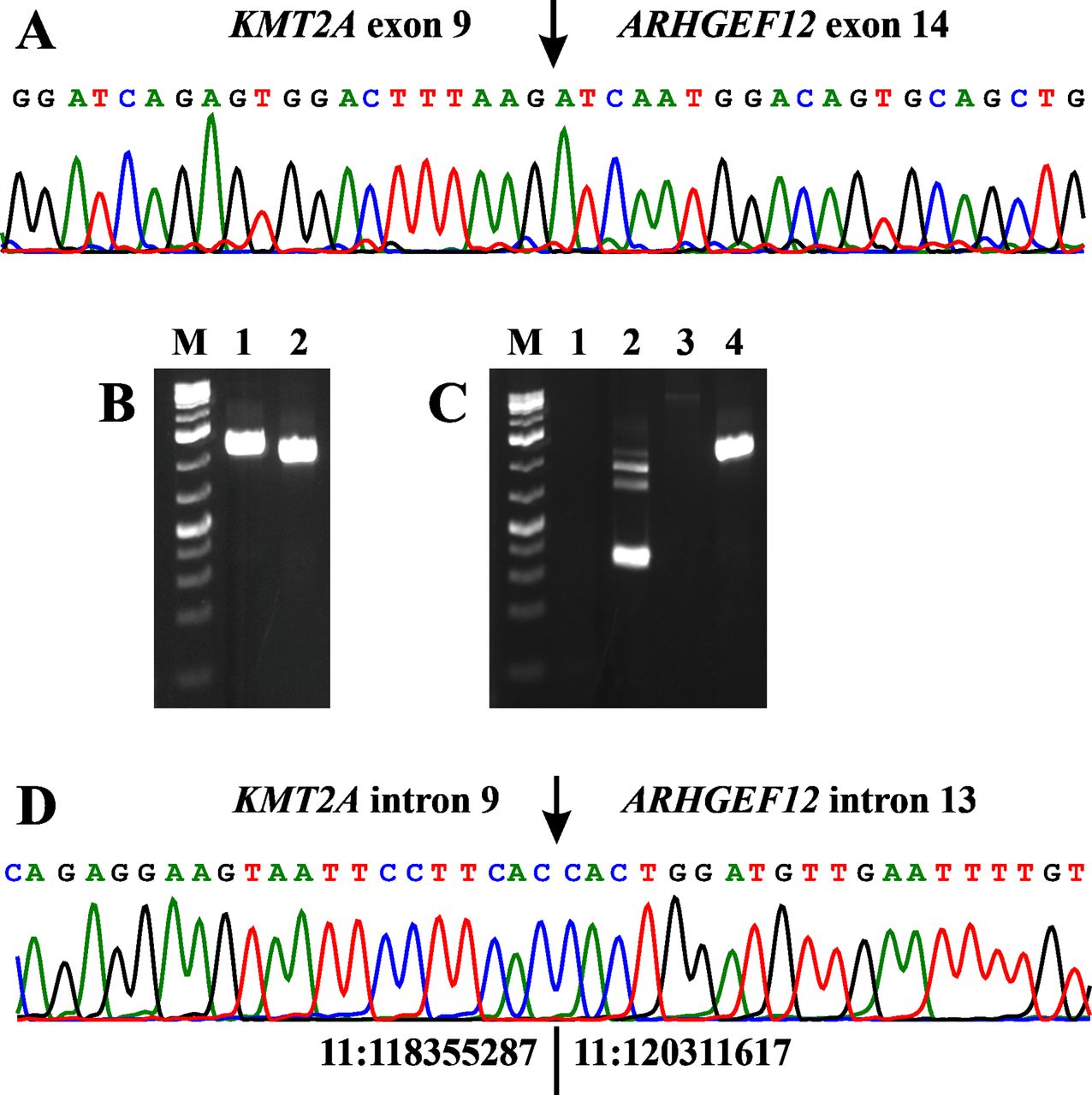
- Figure 3.
PCR analyses of the pediatric leukemias. (A) Partial sequence chromatogram showing the junction position of exon 9 of the KMT2A with exon 14 of ARHGEF12 in the chimeric transcript. (B) Gel electrophoresis showing the amplified KMT2A-ARHGEF12 genomic DNA fragment using two primer combinations and template DNA extracted from the patient’s bone marrow cells 1,539 days after the initial diagnosis. Lane 1: Primer combination MLL-4116F1/ARHGEF12-1502R1. Lane 2: Primer combination MLL-4202-F1/ARHGEF12-1437-R1. (C) Gel electrophoresis showing the absence of amplified KMT2A-ARHGEF12 cDNA (lane 1) and genomic DNA (lane 3) fragments at diagnosis when the leukemic cells had t(9;11)(p21;q23) and a KMT2A-MLLT3 fusion gene, and the presence of KMT2A- ARHGEF12 in the patient’s bone marrow cells 1,198 days after the initial diagnosis, when the patient had developed ALL with t(14;19)(q32;q13) and rearrangement of the IGH locus (lane 4). For both cDNA and genomic DNA amplifications, the primer combination MLL-4116F1/ARHGEF12-1502R1 was used. Lane 2: Assessment of the quality of cDNA synthesis by amplification of a cDNA fragment of ABL1 using the primer combination ABL1-91F1/ABL1-404R1. Lane 4: Amplification of a genomic KMT2A-ARHGEF12 DNA fragment in the patient’s bone marrow cells 1,198 days after the initial diagnosis. M, GeneRuler 1 Kb Plus DNA ladder (ThermoFisher Scientific). (D) Partial sequence chromatogram showing the junction position of intron 9 of the KMT2A with intron 13 of ARHGEF12 in the chimeric amplified DNA fragment. The junction of positions 11:118355287-11:120311617 is based on the human genome GRCh37/hg19 assembly.
- Figure 4.
aCGH showing the deletion in the q arm of chromosome 11. Based on the hg19 assembly, the deletion started from the probe at position Chr11:118355288-118355347 in KMT2A and ended at position Chr11:120290981-120291040 in ARHGEF12. The deletion is approximately 2 Mbp.


In this issue


Jump to section
Related Articles
Cited By...
- Acute Lymphoblastic Leukemia With Near-haploid Karyotype and Philadelphia Chromosome
- Acute Undifferentiated Leukemia With a Balanced t(5;10)(q35;p12) Resulting in Fusion of HNRNPH1 With MLLT10
- Novel MYCBP::EHD2 and RUNX1::ZNF780A Fusion Genes in T-cell Acute Lymphoblastic Leukemia
- Interstitial Deletions Generating Fusion Genes