
Main menu

  • Home
  • Current Issue
  • Archive
  • Info for
    • Authors
    • Advertisers
    • Editorial Board
  • Other Publications
    • Anticancer Research
    • In Vivo
    • Cancer Diagnosis & Prognosis
  • More
    • IIAR
    • Conferences
  • About Us
    • General Policy
    • Contact
  • Other Publications
    • Cancer Genomics & Proteomics
    • Anticancer Research
    • In Vivo

User menu

  • Register
  • Subscribe
  • My alerts
  • Log in
  • My Cart

Search

  • Advanced search
Cancer Genomics & Proteomics
  • Other Publications
    • Cancer Genomics & Proteomics
    • Anticancer Research
    • In Vivo
  • Register
  • Subscribe
  • My alerts
  • Log in
  • My Cart
Cancer Genomics & Proteomics

Advanced Search

  • Home
  • Current Issue
  • Archive
  • Info for
    • Authors
    • Advertisers
    • Editorial Board
  • Other Publications
    • Anticancer Research
    • In Vivo
    • Cancer Diagnosis & Prognosis
  • More
    • IIAR
    • Conferences
  • About Us
    • General Policy
    • Contact
  • Visit iiar on Facebook
  • Follow us on Linkedin
Research Article
Open Access

Transcriptome Alterations in Liver Metastases of Colorectal Cancer After Acquired Resistance to Cetuximab

ZONGCHENG LI, YULING CHEN, WU REN, SHUOFENG HU, ZHAOLI TAN, YAN WANG, YAOWEN CHEN, JIAN ZHANG, JIAQI WU, TINGTING LI, JIANMING XU and XIAOMIN YING
Cancer Genomics & Proteomics May 2019, 16 (3) 207-219; DOI: https://doi.org/10.21873/cgp.20126
ZONGCHENG LI
1Center for Computational Biology, Beijing Institute of Basic Medical Sciences, Beijing, P.R. China
2State Key Laboratory of Proteomics, Translational Medicine Center of Stem Cells, 307-Ivy Translational Medicine Center, Laboratory of Oncology, Affiliated Hospital, Academy of Military Medical Sciences, Beijing, P.R. China
  • Find this author on Google Scholar
  • Find this author on PubMed
  • Search for this author on this site
YULING CHEN
3Department of GI Oncology, 307 Hospital of PLA, Academy of Military Medical Sciences, Beijing, P.R. China
  • Find this author on Google Scholar
  • Find this author on PubMed
  • Search for this author on this site
WU REN
1Center for Computational Biology, Beijing Institute of Basic Medical Sciences, Beijing, P.R. China
4Department of Obstetrics and Gynecology, Tongji Hospital, Tongji Medical College, Huazhong University of Science and Technology, Wuhan, P.R. China
  • Find this author on Google Scholar
  • Find this author on PubMed
  • Search for this author on this site
SHUOFENG HU
1Center for Computational Biology, Beijing Institute of Basic Medical Sciences, Beijing, P.R. China
  • Find this author on Google Scholar
  • Find this author on PubMed
  • Search for this author on this site
ZHAOLI TAN
3Department of GI Oncology, 307 Hospital of PLA, Academy of Military Medical Sciences, Beijing, P.R. China
  • Find this author on Google Scholar
  • Find this author on PubMed
  • Search for this author on this site
YAN WANG
3Department of GI Oncology, 307 Hospital of PLA, Academy of Military Medical Sciences, Beijing, P.R. China
  • Find this author on Google Scholar
  • Find this author on PubMed
  • Search for this author on this site
YAOWEN CHEN
1Center for Computational Biology, Beijing Institute of Basic Medical Sciences, Beijing, P.R. China
5Department of Obstetrics and Gynecology, Fuzhou General Hospital of Nanjing Military Command, Fuzhou, P.R. China
  • Find this author on Google Scholar
  • Find this author on PubMed
  • Search for this author on this site
JIAN ZHANG
1Center for Computational Biology, Beijing Institute of Basic Medical Sciences, Beijing, P.R. China
  • Find this author on Google Scholar
  • Find this author on PubMed
  • Search for this author on this site
JIAQI WU
1Center for Computational Biology, Beijing Institute of Basic Medical Sciences, Beijing, P.R. China
  • Find this author on Google Scholar
  • Find this author on PubMed
  • Search for this author on this site
TINGTING LI
6Department of Geriatric Gastroenterology, Chinese People's Liberation Army General Hospital, Beijing, P.R. China
7State Key Laboratory of Kidney Diseases, Chinese People's Liberation Army General Hospital, Beijing, P.R. China
  • Find this author on Google Scholar
  • Find this author on PubMed
  • Search for this author on this site
JIANMING XU
3Department of GI Oncology, 307 Hospital of PLA, Academy of Military Medical Sciences, Beijing, P.R. China
  • Find this author on Google Scholar
  • Find this author on PubMed
  • Search for this author on this site
  • For correspondence: yingxmbio@foxmail.com yingxm@bmi.ac.cn jmxu2003@yahoo.com
XIAOMIN YING
1Center for Computational Biology, Beijing Institute of Basic Medical Sciences, Beijing, P.R. China
  • Find this author on Google Scholar
  • Find this author on PubMed
  • Search for this author on this site
  • For correspondence: yingxmbio@foxmail.com yingxm@bmi.ac.cn jmxu2003@yahoo.com
  • Article
  • Figures & Data
  • Info & Metrics
  • PDF
Loading

Abstract

Background/Aim: Cetuximab in combination with chemotherapy is recommended as first-line therapy for metastatic colorectal cancer (mCRC) with wild-type RAS. However, drug resistance to cetuximab exists widely in mCRC and reduces the prognosis of patients. Although some genomic alterations have been demonstrated to drive acquired resistance to cetuximab, the overall compendium of inherent molecular mechanisms is still incomplete. Materials and Methods: Four liver metastasis biopsies were collected from two mCRC patients who were treated with cetuximab in combination with 5-fluororacil plus leucovorin and oxaliplatin (FOLFOX) regimen. Results: Transcriptomic analysis revealed global gene expression alterations between paired samples prior to treatment and after acquired resistance. Further bioinformatics analysis discovered differentially expressed protein-coding genes/lncRNAs/miRNAs, potential miRNA-mRNA regulatory networks and lncRNA-mRNA competing endogenous RNA network, which may be potential biomarkers or play roles during the process of acquired resistance to cetuximab. Conclusion: Our study contributes to deciphering the molecular mechanisms of acquired resistance to cetuximab.

  • Acquired resistance
  • cetuximab
  • colorectal cancer
  • transcriptome alteration
  • regulatory networks

Colorectal carcinoma (CRC) is one of the most common cancers and has the third leading incidence rate worldwide. About 1.2 million people are newly diagnosed with colorectal cancer per year (1). Distant metastasis is the main reason for death in CRC patients. About 25% of CRC patients have distant metastasis at first diagnosis, and 50% of stage I to stage III patients will eventually develop metastasis (2). Liver is the main target organ of metastasis in colorectal cancer (mCRC), and the majority of CRC patients with liver metastases are unable to get radical resection. For these patients, the main purpose of treatment is to prolong survival, prevent further tumor progression, reduce tumor-related symptoms and improve quality of life (2).

In recent years, the application of chemotherapy and molecular targeted therapies, including anti-epidermal growth factor receptor (EGFR) monoclonal antibodies and anti-vascular endothelial growth factor (VEGF) monoclonal antibodies, has improved the prognosis of mCRC patients. The National Cancer Institute lists 24 drugs for the treatment of mCRC (see list in http://www.cancer.gov/about-cancer/treatment/drugs/colorectal), among which cetuximab is one of the most effective monoclonal antibodies targeting EGFR on the cell membrane (3) and can significantly improve the prognosis of mCRC patients with wild-type RAS (4-9). Cetuximab in combination with chemotherapy has been recommended as first-line therapy (10).

However, drug resistance to cetuximab exists widely. Clinical studies show that cetuximab alone is effective for only about 10% of mCRC patients (7). Many efforts have been made to find biomarkers or drivers of drug resistance to cetuximab, in order to increase the effectiveness of cetuximab and improve the treatment of mCRC. Drug resistance to cetuximab can be classified into primary and secondary resistance (i.e. acquired resistance). The main causes of primary drug resistance are mutations in codons 12 and 13 at the exon 2 of the KRAS gene (6, 11-13), which has been used for routine testing and guiding the clinical treatment (8, 10). Moreover, several other genomic alteration events, including mutations in KRAS exons 3 and 4, NRAS exon 2, 3 and 4, BRAF V600E, PIK3CA and HER2 amplification, have also been reported to be associated with primary drug resistance to cetuximab (14-18). Acquired resistance to cetuximab often occurs at 3 to 12 months after effective response to treatment (7). Mutations in KRAS and NRAS genes are the most common causes for acquired resistance to cetuximab (19, 20). The amplification of HER2 and MET genes, the other two members of the receptor tyrosine kinases (RTK), can also lead to acquired resistance by activating downstream RAF-MEK-ERK signaling pathway (21, 22). In addition, the S492R mutation in the extracellular domain of EGFR can also result in acquired resistance by hindering antibodies from binding to EGFR (23).

Although some genomic alterations have been identified and demonstrated to drive acquired resistance to cetuximab, the overall compendium of inherent molecular mechanisms is still incomplete. Transcriptomic analysis can provide comprehensive insights into molecular mechanisms, which include differential expression analysis and pathway/regulation networks of protein-coding genes, long non-coding RNAs (lncRNA) and miRNAs. However, transcriptome alterations, especially alterations between matched biopsies prior to treatment and after acquired resistance, are largely unknown up to date.

In this study, we collected four liver metastasis biopsies from two mCRC patients who were treated with cetuximab in combination with 5-fluororacil plus leucovorin and oxaliplatin (FOLFOX). Each patient had undergone ultrasound-guided biopsies prior to treatment and after acquired resistance (tumor re-progression after effective response to treatment for more than six months). High-throughput transcriptome sequencing, including RNA-Seq and small RNA-Seq, were conducted for all the four samples. Transcriptomic analysis revealed gene expression alterations between paired samples prior to treatment and after acquired resistance. Further bioinformatics analysis discovered differentially expressed protein-coding genes/lncRNAs/miRNAs, potential miRNA-mRNA regulatory networks and lncRNA-mRNA competing endogenous RNA (ceRNA) network, which may be potential biomarkers or play roles during the process of acquired resistance to cetuximab. Our study may contribute to deciphering the molecular mechanisms of acquired resistance to cetuximab.

Materials and Methods

Patient inclusion and sample collection. A retrospective single-center study was performed at the 307 Hospital, Academy of Military Medical Sciences, Beijing, China. Eligible patients had pathologically confirmed metastatic colorectal cancer (mCRC) harboring wild-type KRAS codons 12 and 13 and BRAF codon 600 determined via Sanger sequencing of the tumor tissue DNA, and they received targeted therapy and chemotherapy for the first time. Patients received cetuximab in combination with FOLOX chemotherapy. Computed tomography (CT) scans were performed and reviewed every 4 to 6 weeks to evaluate clinical response using the Response Evaluation Criteria in Solid Tumors (RECIST), version 1.1 (24). All the patients signed an informed consent for liver biopsy and publication of the results. Fresh tumor tissues from metastatic liver lesions were obtained at baseline and disease progression. Tumor biopsies were frozen at liquid nitrogen immediately when taken out and were linked to demographic, clinical and genetic data stored in a secure research database. This study protocol was approved by the local ethics committee (KY-2011-8-3). Tissue acquisition and handling of human tissue specimens were carried out in accordance with the institutional and state regulations.

Treatment of cetuximab in combination with chemotherapy. All of these agents were administered intravenous (IV) via an ambulatory pump and a central venous line. Treatment was continued until disease progression. Cetuximab: 400 mg/m2 IV over 2 h at the first week as an initial dose, then 250 mg/m2 IV over 1 h weekly. FOLFOX regimen: Oxaliplatin 85 mg/m2, IV over 2 h, day 1; Leucovorin 400 mg/m2, IV over 2 h, day 1; 5-fluorouracil 400 mg/m2, bolus on day 1, then 2,400 mg/m2 IV infusion over 46 h.

Liver biopsy and pathology assessment. Ultrasound guided liver biopsies were performed using a 18 gauge biopsy needle, 2 cores with a minimum length greater than 5 mm were taken, and each of the cores were mirror sectioned, half of which were send to pathology department for H&E staining and pathological evaluation to ensure the tumor content was more than 50%. The rest of the tissue was placed in a freezing tube with RNAlater, frozen at −80°C.

RNA isolation, library construction and RNA sequencing. Total RNA was isolated from fresh frozen tumor tissues using Trizol reagent. Library constructions of Poly-A mRNA and small RNA were conducted using TruSeq Stranded mRNA Library Prep Kit (Illumina, San Diego, CA, USA) and TruSeq Small RNA Library Preparation Kits (Illumina, USA). The high-throughput sequencing was carried out using Illumina Hiseq 2000.

Mapping and quantification. For mRNA sequencing data, poor quality reads were filtered from the raw paired-end reads using custom perl script. Then the paired-end reads were aligned to the hg19 human genome assembly using Tophat program version 2.0.13 (25). The resulting alignments were saved as a BAM file. The raw counts for each gene were calculated using the HTSeq-count script (26) by counting the number of reads overlapping with genes (mRNAs and lncRNAs) annotated in GENCODE version 15 (27). For small RNA sequencing data, after removing adaptors and poor quality reads, small RNA reads were aligned to know human miRNAs from miRBase v21 (28). The raw count for each miRNA was calculated. The workflow is illustrated in Supplementary Figure 1.

Figure 1.
  • Download figure
  • Open in new tab
  • Download powerpoint
Figure 1.

Pathological sections and CT scans of liver lesions. (A) H&E staining picture of primary tumor of colon (left) and liver metastasis (right). (B) Tumor response to cetuximab containing therapy in patient mCRC002 with liver metastases. CT scan revealed multiple heterogeneously enhancing tumor masses in the right and left lobes of liver, after 2 cycles (8 weeks) of chemotherapy plus Cetuximab, dramatic tumor regression at all sites of disease were seen, the regression lasted 24 weeks. After continued therapy for more than 7 months, the patient developed disease progression.

Estimation of tumor purity. Tumor purity of the four samples was estimated using the ESTIMATE R package (29). ESTIMATE method uses gene expression profiles of 141 immune genes and 141 stromal genes to obtain ESTIMATE scores and purity was estimated using the formula described in Yoshihara et al. (29).

Categories and subcellular localization of protein-coding genes. Significantly up-regulated genes after acquired resistance were assigned to different categories and subcellular localizations according to Uniprot database (30).

Literature search. The combination of gene name and any of these keywords, including “cancer”, “tumor”, “resistant”, “resistance”, “anti-drug” and “drug-resistance”, were used in PubMed literature search. According to the results of the literature search, differentially expressed genes were manually classified into five groups, which are tumor-related genes (“cancer” and “tumor”), drug resistance-related genes (“resistant”, “resistance”, “anti-drug” and “drug-resistance”), drug resistance genes (“resistant”, “resistance”, “anti-drug” and “drug-resistance”), tumor suppressor genes (“cancer” and “tumor”) and other genes.

Construction of miRNA-mRNA negative regulatory network and ceRNA network. miRwalk (31), at default settings, was used to predict target mRNAs for each differentially expressed miRNA. The potential target mRNAs, which were supported by at least three prediction methods of miRwalk, RNA22, miRANDA and Targetscan and differentially expressed in opposite direction of the miRNA, were retained. Then, these potential regulatory links between miRNAs and mRNAs with significant opposite expression were used for construction of a miRNA-mRNA negative regulatory network.

Based on gene annotations of GENCODE v15 (27), we selected lncRNAs with the given “gene_type”, including lincRNA, antisense RNA, sense intronic, overlap sense and processed_transcript. The ceRNA prediction procedure includes four steps (Supplementary Figure 2): i) predicting the lncRNA-miRNA interactions based on the CLIP-Seq data from Starbase database (Version 2.0) for differentially expressed lncRNA; ii) obtaining experiment validated miRNA-mRNA pairs based on the miRtarbase database for differentially expressed mRNAs; iii) generating potential lncRNA-mRNA relationships connected by miRNAs; iv) excluding lncRNA-mRNA pairs which showed expression changes in different directions. Cytoscape was employed for visualization of networks.

Availability of data and material. Raw data and gene expression profiles for this study are accessible via the GEO repository (http://www.ncbi.nlm.nih.gov/geo/) under accession identification number GSE84270. Supplementary tables and supplementary figures can be downloaded from http://www.omicsnet.org/pub/coloncancer.html.

Statistical analysis. For gene or miRNA differential expression analysis, paired sample design was employed using R package DESeq2 version 1.8.0 (32), which was a powerful method for the detection of differentially expressed genes of count data from RNA-Seq using shrinkage estimation for dispersions and fold changes to improve stability and interpretability of estimates. Fold change >1.5 and FDR <0.05 was adopted as a statistically significant threshold. The variance Stabilizing Transformation function was used to normalize the count data for visualization.

Results

Clinical and treatment information of patients. Two patients who were newly diagnosed and pathologically confirmed with metastatic colorectal cancer (mCRC) harboring wild-type KRAS codons 12 and 13 and BRAF codon 600 were screened for eligibility between August 2011 and December 2013. They were treated with cetuximab in combination with FOLFOX regimen (see Materials and Methods) and obtained continuous partial responses for more than six months. CT scans of liver lesions were performed every four to six weeks. The scans at baseline, best response and disease progression are shown in Figure 1. Detailed clinical and treatment information were provided in Supplementary Table I.

Transcriptome expression profiles of four mCRC biopsies. The four biopsy samples were evaluated by two pathologists and tumor purities were confirmed to be greater than 50%. Each biopsy sample was subjected to high-throughput RNA and small RNA sequencing to obtain expression profiles of genes (protein-coding genes and lncRNAs) and miRNAs. We evaluated the tumor purity scores using ESTIMATE algorithm (29) and gene expression profiles. The tumor purity scores of the four samples ranged from 0.79 to 0.92 (Supplementary Table II), ensuring the reliability of the analysis results. Next, we compared gene and miRNA expression levels between paired samples of prior treatment and after acquired resistance for each patient (Figure 2). The results showed that many genes (Figure 2A and B) and a few miRNAs (Figure 2C and D) were significantly up- or down-regulated in the two patients after drug resistance occurred.

Differentially expressed protein-coding genes after acquired resistance to cetuximab. We identified 782 protein-coding genes differentially expressed between biopsy samples of prior treatment and after acquired resistance. Among them, 699 genes were up-regulated in the biopsy samples after acquired resistance and 83 genes were down-regulated (Figure 3A, Supplementary Table III). The up-regulated gene number is eight times greater than that of the down-regulated genes. Moreover, the change folds of up-regulated genes are much greater than those of the down-regulated genes (11.17±0.64 vs. 5.23±0.29, p<0.001, Mann-Whitney U-test). Specifically, 282 genes were up-regulated by >10 fold and an adjusted p-value <0.01, covering 40.3% (282/699) of all the up-regulated genes, whereas only 5 genes were down-regulated by >10 fold and adjusted p value <0.01, covering 6.0% of all the down-regulated genes. These data show that many more genes were activated than repressed when resistance occurred.

GO annotation and KEGG pathway enrichment analysis was then performed on 699 significantly up-regulated genes and 83 down-regulated genes using DAVID webserver (33, 34), respectively. The top five up-regulated biological processes were: small molecule metabolic process, oxidation-reduction process, xenobiotic metabolic process, regulation of complement activation, and negative regulation of endopeptidase activity. The top five enriched up-regulated KEGG pathways were: complement and coagulation cascades, metabolic pathways, chemical carcinogenesis, glycine, serine and threonine metabolism, and retinol metabolism (Figure 3B). These results showed that metabolism and complement activity are more active after resistance to cetuximab occurred, which is consistent with the previous reports showing that metabolic alterations help cancer cells to proliferate rapidly and develop drug resistance phenotypes (35, 36). However, the down-regulated genes had no enriched biological processes or KEGG pathways.

Four keratins, KRT1, KRT14, KRT5 and KRT2, were most significantly up-regulated among all the up-regulated genes (Supplementary Table III). The change folds were all greater than 110, far greater than those of other up-regulated genes. KRT1, KRT2 and KRT5 are type II cytokeratins and KRT14 is a type I cytokeratin. KRT5 and KRT14 are usually expressed in the basal layer of the epidermis and stratified squamous epithelia, and form heterotetramers. KRT5 and KRT14 are diagnostic markers in breast cancer (37), and their high expression indicates aggressive behavior and poor prognosis (38, 39). KRT1 is a diagnostic marker in skin squamous cancer (37). Our result suggests that KRT1, KRT2, KRT5 and KRT14 may be markers of resistance to cetuximab in mCRC.

Figure 2.
  • Download figure
  • Open in new tab
  • Download powerpoint
Figure 2.

Comparisons of gene and miRNA expression between paired samples of prior treatment and after acquired resistance for each patient. Scatterplots of gene and miRNA expression between paired samples prior to treatment and after acquired resistance for mCRC001 (A for genes and B for miRNAs) and mCRC002 (C for genes and D for miRNAs). Grey line stands for y=x; red line stands for y=x+2; green line stands for y=x−2. In panel A for example, black points above red line account for genes up-regulated more than twice in the acquired resistance sample, while black points under green line account for genes down-regulated more than twice in the acquired resistance sample. Histograms alongside each scatterplot show distributions of gene and miRNA expression of corresponding samples (cyan is for sample of prior treatment and brown for acquired resistance).

We further inspected the kinases, cytokines and cell surface receptors in the up-regulated genes (Figure 3C), since they can more readily become drug targets. Two nuclear receptors, ESR1 (Estrogen receptor 1) and AR (Androgen receptor), were up-regulated after acquired resistance to cetuximab. Six kinases were also up-regulated, including MET, RET, MST1, PDK4, KHK and DAPK1. Eight cell surface receptors were up-regulated, including MET, RET, GHR, IL1R1, IL6R, SMO, GFRA1 and GFRA2. Twelve growth factors and cytokines were up-regulated, including CCL16, CCL19, CCL21, CXCL12, FGF21, GDF2, GREM2, IGF1, IGF2, INHBC, INHBE and NRG1. Among these genes, MET and RET are members of the RTK (Receptor tyrosine kinase) family (40), the same as EGFR which is the target of cetuximab. Amplification of MET gene has been reported to lead to acquired resistance to cetuximab (21, 22). RET (41) and ESR1 (42, 43) were reported to correlate with endocrine resistance in breast cancer. SMO gene amplification was associated with resistance to EGFR TKIs in human lung cancer (44). NGR1 was reported to provide resistance to MEK inhibitors in metastatic uveal melanoma (45). Our results suggested that these up-regulated kinases, cytokines and cell surface receptors may play roles in acquired resistance to cetuximab and that the inhibitors or drugs targeting these proteins may sensitize CRC to cetuximab treatment.

A literature search was also conducted for all 699 up-regulated genes (see Materials and methods). Twenty-one genes have been reported to lead to drug resistance in cancers (Figure 3D, Supplementary Table IV). Fifty-six genes have been shown to correlate with drug resistance, sixty-two genes are known cancer genes and 171 genes have been reported to be associated with cancer (Figure 3D, Supplementary Table III). This result showed that nearly half (296/699=42.3%, Supplementary Table III) of the up-regulated genes are known drug resistance or cancer associated genes. The rest of the genes may also play roles in drug resistance or cancer development and are worth further validation.

Differentially expressed lncRNAs after acquired resistance to Cetuximab. Fifty-three lncRNAs were differentially expressed between biopsies of prior treatment and after acquired resistance (25 up-regulated and 28 down-regulated, Supplementary Table III). The change folds of up-regulated lncRNAs were much greater than those of down-regulated lncRNAs (10.82±1.15 vs. 4.97±0.32, p<0.001, Mann-Whitney U-test). The top two significantly up-regulated lncRNA were H19 [fold change (FC)=16.7, adjusted p<0.001] and HULC (FC=12.5, adjusted p<0.001, Supplementary Table III). Up-regulation of H19 was reported to be associated with drug resistance in human hepatocellular carcinoma cells (46). Up-regulation of HULC was reported to promote the progression of hepatocellular carcinoma (47) and CRC (48). Two down-regulated lncRNAs MALAT1 (FC=3.7, adjusted p=0.005) and GAS5 (FC=3.3, adjusted p=0.047, Supplementary Table III) were reported to be associated with drug resistance. Down-regulation of MALAT1 was observed in vincristine-resistant acute lymphoblastic leukemia (49). Down-regulation of GAS5 leads to trastuzumab resistance in breast cancer (50) and was reported to be associated with cisplatin resistance in non-small cell lung cancer (NSCLC) (51). Our results suggest that these differentially expressed lncRNAs may also play roles in resistance to cetuximab in mCRC.

Interestingly, more lncRNAs are down-regulated in acquired resistance samples even though down-regulated genes were significantly less than up-regulated genes (p<0.001, one-sided Fisher's Exact Test, Supplementary Table V). This result indicates that high expression of these down-regulated lncRNAs may play important roles in sensitizing CRC to cetuximab treatment.

Potential miRNA regulatory network and competitive endogenous RNAs associated with Cetuximab resistance. Twenty mature miRNAs were significantly up-regulated (>3 folds) and nine mature miRNAs were significantly down-regulated (>3 folds) when resistance to cetuximab occurred (Figure 4A, Supplementary Table VI). The 5’ and 3’ cleavage products (denoted as -5p and -3p in the names of mature miRNAs) of four miRNA precursors were both up-regulated in biopsies after acquired resistance, including mir-885, mir-122, mir-483, and mir-139. (Supplementary Table VI). Interestingly, miR-885-5p was reported to be up-regulated in non-small cell lung cancer cell resistance to gemcitabine (52), and was up-regulated in liver metastasis specimens of CRC patients (53). miR-885-5p was the most up-regulated miRNA in lymphovascular invasion carcinoid tumors compared with tumors without invasion (54). Even more interestingly, miR-885-5p was detected in serum. High serum miR-885-5p expression independently predicted prognosis and metastasis in CRC (55). These results suggest that the significantly up- or down-regulated miRNAs may potentially become biomarkers for cetuximab resistance.

We further built the miRNA-mRNA negative regulatory network among differentially expressed miRNAs and mRNAs (Figure 4B, C, Supplementary Figure 1). Although down-regulated miRNAs were much less in number than up-regulated miRNAs (9 vs. 20), the target mRNAs of the down-regulated miRNAs were much more than those of the up-regulated miRNAs (Average target mRNA number: 35.22±22.16 vs. 5.53±3.45, p=0.004, t-test). These results suggest that the down-regulated miRNAs may play more important roles in resistance to cetuximab in mCRC. Up-regulation of the down-regulated miRNAs might sensitize CRC to cetuximab treatment.

Competitive endogenous RNA (ceRNA) is a pair of RNAs, which have the same miRNA response element (MRE) and may competing for the same endogenous miRNAs, and their expression changes in the same direction (56, 57). Based on this assumption, we predicted the ceRNA network among differentially expressed mRNAs and lncRNAs (Figure 4D). Seven lncRNAs and 32 mRNAs formed the ceRNA network. H19, the top significantly up-regulated lncRNAs, had the most potentially competitive mRNAs. The other 6 lncRNAs had 1 to 8 potentially competitive mRNAs. Some lncRNAs and mRNAs with known functions in cancer were involved in the network. For example, lncRNA H19, an imprinted gene, plays an important role in mammalian development (58) and has been reported to be related to development, metastasis and drug-resistance of various tumors (59-61). LncRNA Gas5, a tumor suppressor, has been correlated with drug resistance (62, 63). MMP9, a member of the MMP family, plays an important role in early progression of tumor and may promote proliferation, adhesion, migration and angiogenesis (64-66). CDKN2A (P16) is a known tumor suppressor gene (67-69). WIF1 is known to inhibit the Wnt protein by interacting with Frizzled, and then inhibit Wnt/β-catenin signaling transduction (70-72). The competitive relationship between these lncRNAs and mRNAs may exert functions in tumor resistance to cetuximab.

Figure 3.
  • Download figure
  • Open in new tab
  • Download powerpoint
Figure 3.

Analysis of differentially expressed genes after acquired resistance to cetuximab. (A) Heatmap of 835 differentially expressed genes between 2 groups prior to treatment and after acquired resistance. (B) The results of the gene functional enrichment analysis of 699 up-regulated protein-coding genes. Top panel shows the top 5 enriched KEGG pathways, and bottom panel shows the top 5 enriched biological processes. (C) Subcellular components and location of 26 specific genes, which code for cytokine, kinase, and cell surface receptors, among up-regulated genes. (D) Pie chart of categories of up-regulated genes identified by literature search.

Figure 4.
  • Download figure
  • Open in new tab
  • Download powerpoint
Figure 4.

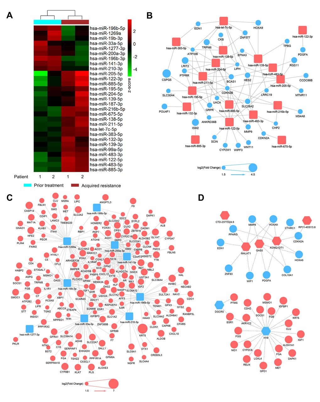
Integrative analysis of differentially expressed miRNAs, mRNAs and lncRNAs identified potential regulatory networks. (A) Heatmap of 29 differentially expressed miRNAs between 2 groups prior to treatment and after acquired resistance. (B and C) Predicted miRNA-mRNA negative regulatory network among differentially expressed miRNAs and mRNAs. Yellow squares represent up-regulated miRNAs (B) and down-regulated miRNAs (C). Circles represent down-regulated (blue in B) and up-regulated (red in C) mRNAs, and circle sizes are proportional to fold changes. (D) Predicted lncRNA-mRNA ceRNA regulatory network among differentially expressed lncRNAs and mRNAs. Yellow squares represent down-regulated (left panel) and up-regulated lncRNAs (right panel). Circles represent up-regulated (red in left panel) and down-regulated (blue in right panel) mRNAs.

Discussion

Acquired resistance to cetuximab widely occurs in the clinic. However, since paired biopsies prior to treatment and after acquired resistance from mCRC patients can rarely be obtained, global transcriptome alterations of paired biopsies are still unknown. We obtained four paired biopsy samples from two liver mCRCs, prior treatment with cetuximab and 5-FOLOX and after acquired resistance. By using high-throughput RNA and small RNA sequencing technology, we delineated global transcriptome changes when resistance occurred. Differential expression analysis, miRNA regulatory network and ceRNA network analysis suggested that some protein-coding genes, lncRNAs and miRNAs may be biomarkers and may play roles in the acquired resistance to cetuximab in mCRC. Our results provide clues for further studies of the molecular mechanisms of acquired resistance to cetuximab.

Cetuximab targets EGFR on the cell membrane, which is a member of the RTK family. The mutations or amplifications of several RTK family genes, including KRAS, NRAS, HER2 and MET, have been reported to result in acquired resistance to cetuximab (19-23). In our analysis, another RTK family member, RET, was significantly up-regulated in the biopsies of acquired resistance. Furthermore, three miRNAs miR-196b-5p, miR-141-3p and miR-200a-3p predicted to target RET mRNA were down-regulated in the biopsies of acquired resistance. RET has been reported to be related to drug resistance to endocrine therapy (73). Our results suggest that up-regulation of RET might also cause cetuximab resistance in mCRC.

Twenty-nine miRNAs were significantly up- or down-regulated in the biopsies of acquired resistance. MiRNAs are small molecules, ~22 nt in length. Some miRNAs can be detected in serum, for example, miR-885-5p. In our analysis, miR-885-5p was up-regulated in acquired resistance samples. It was reported that high serum miR-885-5p expression independently predicted prognosis and metastasis in CRC (55). These results suggest that miR-885-5p might be a serum biomarker of acquired resistance to cetuximab.

The functions and mechanisms of miRNAs and lncRNAs in acquired resistance are largely unknown. By constructing miRNA-mRNA regulatory networks and lncRNA-mRNA ceRNA networks, we predicted potential regulatory mechanisms for miRNAs and lncRNAs. For example, several miRNAs such as miR-141-3p and miR-1269a might contribute to sensitizing CRC to cetuximab treatment by inhibiting the genes that drive acquired resistance. LncRNAs such as H19 and MALAT1 might regulate critical genes that drive or inhibit resistance as competitive endogenous RNAs. This information provides clues for investigating the roles and mechanisms of miRNAs and lncRNAs in resistance to cetuximab.

Taken together, we depicted global transcriptome alterations between paired biopsy samples prior to treatment and after acquired resistance from two liver mCRCs treated with cetuximab and 5-FOLOX. To our knowledge, this is the first transcriptome profiling for paired biopsies of prior treatment and acquired resistance to cetuximab. The conclusion is preliminary due to the small sample size. Nonetheless, our results still provide clues for subsequent studies on the biomarkers and molecular mechanisms of cetuximab resistance.

Conclusion

Our study contributes to deciphering the molecular mechanisms of acquired resistance to cetuximab.

Acknowledgements

The Authors would like to thank all the medical and ancillary staff at the Cancer Center and the patients for consenting to participate. The Authors also thank Yun Shao and Yanhong Tai of the Department of Pathology for assistance with the preparation and assessment of formalin-fixed paraffin-embedded tumor samples, Gong-Jie Li of the Department of Radiology for assistance with radiological response evaluation and Tao Li and Xin Pan for their helpful discussions.

Footnotes

  • ↵* These Authors contributed equally to this work.

  • Authors' Contributions

    J. X. and X.Y. conceived the study; W.R., Z.L. and X.Y. wrote the manuscript; Z.L. and W.R. performed the transcriptome analysis with the help of S.H., Y.C., J.Z. and J.W.; Y.C., Y. W., Z. T. and T. L. conducted the clinical sample preparation and high-throughput sequencing. X. Y. and J. X. revised the manuscript. All authors reviewed the manuscript.

  • This article is freely accessible online.

  • Conflicts of Interest

    The Authors declare no competing financial interests.

  • Funding

    This work was supported by National Key R&D Program of China (Grant No. 2017YFC0908300 to X. Ying), the China National High Technology Research and Development Program (No. 2014AA020604 to X. Ying), the National Natural Science Foundation of China Project (No. 81573458 to J. Xu), and the National Natural Science Foundation of China (Grant No. 81703006 to T. Li).

  • Received February 17, 2019.
  • Revision received April 5, 2019.
  • Accepted April 8, 2019.
  • Copyright© 2019, International Institute of Anticancer Research (Dr. George J. Delinasios), All rights reserved

References

  1. ↵
    1. Brenner H,
    2. Kloor M,
    3. Pox CP
    : Colorectal cancer. Lancet Lond Engl 383: 1490-1502, 2014. PMID: 24225001. DOI: 10.1016/S0140-6736(13)61649-9
    OpenUrl
  2. ↵
    1. Van Cutsem E,
    2. Cervantes A,
    3. Nordlinger B,
    4. Arnold D,
    5. ESMO Guidelines Working Group
    : Metastatic colorectal cancer: ESMO Clinical Practice Guidelines for diagnosis, treatment and follow-up. Ann Oncol 25: iii1-9, 2014. PMID: 25190710. DOI: 10.1093/annonc/mdu260
    OpenUrlCrossRefPubMed
  3. ↵
    1. Giampieri R,
    2. Scartozzi M,
    3. Prete MD,
    4. Maccaroni E,
    5. Bittoni A,
    6. Faloppi L,
    7. Bianconi M,
    8. Cecchini L,
    9. Cascinu S
    : Molecular biomarkers of resistance to anti-EGFR treatment in metastatic colorectal cancer, from classical to innovation. Crit Rev Oncol Hematol 88: 272-283, 2013. PMID: 23806981. DOI: 10.1016/j.critrevonc.2013.05.008
    OpenUrl
  4. ↵
    1. Ciardiello F,
    2. Tortora G
    : EGFR Antagonists in Cancer Treatment. N Engl J Med 358: 1160-1174, 2008. PMID: 18337605. DOI: 10.1056/NEJMra0707704
    OpenUrlCrossRefPubMed
    1. Jonker DJ,
    2. O'Callaghan CJ,
    3. Karapetis CS,
    4. Zalcberg JR,
    5. Tu D,
    6. Au H-J,
    7. Berry SR,
    8. Krahn M,
    9. Price T,
    10. Simes RJ,
    11. Tebbutt NC,
    12. van Hazel G,
    13. Wierzbicki R,
    14. Langer C,
    15. Moore MJ
    : Cetuximab for the treatment of colorectal cancer. N Engl J Med 357: 2040-2048, 2007. PMID: 18003960. DOI: 10.1056/NEJMoa071834
    OpenUrlCrossRefPubMed
  5. ↵
    1. Karapetis CS,
    2. Khambata-Ford S,
    3. Jonker DJ,
    4. O'Callaghan CJ,
    5. Tu D,
    6. Tebbutt NC,
    7. Simes RJ,
    8. Chalchal H,
    9. Shapiro JD,
    10. Robitaille S,
    11. Price TJ,
    12. Shepherd L,
    13. Au HJ,
    14. Langer C,
    15. Moore MJ,
    16. Zalcberg JR
    : K-ras mutations and benefit from cetuximab in advanced colorectal cancer. N Engl J Med 359: 1757-1765, 2008. PMID: 18946061. DOI: 10.1056/NEJMoa0804385
    OpenUrlCrossRefPubMed
  6. ↵
    1. Cunningham D,
    2. Humblet Y,
    3. Siena S,
    4. Khayat D,
    5. Bleiberg H,
    6. Santoro A,
    7. Bets D,
    8. Mueser M,
    9. Harstrick A,
    10. Verslype C,
    11. Chau I,
    12. Van Cutsem E
    : Cetuximab monotherapy and cetuximab plus irinotecan in irinotecan-refractory metastatic colorectal cancer. N Engl J Med 351: 337-345, 2004. PMID: 15269313. DOI: 10.1056/NEJMoa033025
    OpenUrlCrossRefPubMed
  7. ↵
    1. Van Cutsem E,
    2. Köhne CH,
    3. Hitre E,
    4. Zaluski J,
    5. Chang Chien CR,
    6. Makhson A,
    7. D'Haens G,
    8. Pintér T,
    9. Lim R,
    10. Bodoky G,
    11. Roh JK,
    12. Folprecht G,
    13. Ruff P,
    14. Stroh C,
    15. Tejpar S,
    16. Schlichting M,
    17. Nippgen J,
    18. Rougier P
    : Cetuximab and chemotherapy as initial treatment for metastatic colorectal cancer. N Engl J Med 360: 1408-1417, 2009. PMID: 19339720. DOI: 10.1056/NEJM oa0805019
    OpenUrlCrossRefPubMed
  8. ↵
    1. Van Cutsem E,
    2. Peeters M,
    3. Siena S,
    4. Humblet Y,
    5. Hendlisz A,
    6. Neyns B,
    7. Canon JL,
    8. Van Laethem JL,
    9. Maurel J,
    10. Richardson G,
    11. Wolf M,
    12. Amado RG
    : Open-label phase III trial of panitumumab plus best supportive care compared with best supportive care alone in patients with chemotherapy-refractory metastatic colorectal cancer. J Clin Oncol Off J Am Soc Clin Oncol 25: 1658-1664, 2007. PMID: 17470858. DOI: 10.1200/ JCO.2006.08.1620
    OpenUrl
  9. ↵
    1. Bokemeyer C,
    2. Bondarenko I,
    3. Makhson A,
    4. Hartmann JT,
    5. Aparicio J,
    6. Braud F de,
    7. Donea S,
    8. Ludwig H,
    9. Schuch G,
    10. Stroh C,
    11. Loos AH,
    12. Zubel A,
    13. Koralewski P
    : Fluorouracil, leucovorin, and oxaliplatin with and without cetuximab in the first-line treatment of metastatic colorectal cancer. J Clin Oncol 27: 663-671, 2009. PMID: 19114683. DOI: 10.1200/JCO.2008.20.8397
    OpenUrlAbstract/FREE Full Text
  10. ↵
    1. Lièvre A,
    2. Bachet JB,
    3. Le Corre D,
    4. Boige V,
    5. Landi B,
    6. Emile JF,
    7. Côté JF,
    8. Tomasic G,
    9. Penna C,
    10. Ducreux M,
    11. Rougier P,
    12. Penault-Llorca F,
    13. Laurent-Puig P
    : KRAS mutation status is predictive of response to cetuximab therapy in colorectal cancer. Cancer Res 66: 3992-3995, 2006. PMID: 16618717. DOI: 10.1158/0008-5472.CAN-06-0191
    OpenUrlAbstract/FREE Full Text
    1. Benvenuti S,
    2. Sartore-Bianchi A,
    3. Di Nicolantonio F,
    4. Zanon C,
    5. Moroni M,
    6. Veronese S,
    7. Siena S,
    8. Bardelli A
    : Oncogenic activation of the RAS/RAF signaling pathway impairs the response of metastatic colorectal cancers to anti-epidermal growth factor receptor antibody therapies. Cancer Res 67: 2643-2648, 2007. PMID: 17363584. DOI: 10.1158/0008-5472.CAN-06-4158
    OpenUrlAbstract/FREE Full Text
  11. ↵
    1. Amado RG,
    2. Wolf M,
    3. Peeters M,
    4. Van Cutsem E,
    5. Siena S,
    6. Freeman DJ,
    7. Juan T,
    8. Sikorski R,
    9. Suggs S,
    10. Radinsky R,
    11. Patterson SD,
    12. Chang DD
    : Wild-type KRAS is required for panitumumab efficacy in patients with metastatic colorectal cancer. J Clin Oncol 26: 1626-1634, 2008. PMID: 18316791. DOI: 10.1200/JCO.2007.14.7116
    OpenUrlAbstract/FREE Full Text
  12. ↵
    1. Loupakis F,
    2. Ruzzo A,
    3. Cremolini C,
    4. Vincenzi B,
    5. Salvatore L,
    6. Santini D,
    7. Masi G,
    8. Stasi I,
    9. Canestrari E,
    10. Rulli E,
    11. Floriani I,
    12. Bencardino K,
    13. Galluccio N,
    14. Catalano V,
    15. Tonini G,
    16. Magnani M,
    17. Fontanini G,
    18. Basolo F,
    19. Falcone A,
    20. Graziano F
    : KRAS codon 61, 146 and BRAF mutations predict resistance to cetuximab plus irinotecan in KRAS codon 12 and 13 wild-type metastatic colorectal cancer. Br J Cancer 101: 715-721, 2009. PMID: 19603018. DOI: 10.1038/sj.bjc.6605177
    OpenUrlCrossRefPubMed
    1. De Roock W,
    2. Claes B,
    3. Bernasconi D,
    4. De Schutter J,
    5. Biesmans B,
    6. Fountzilas G,
    7. Kalogeras KT,
    8. Kotoula V,
    9. Papamichael D,
    10. Laurent-Puig P,
    11. Penault-Llorca F,
    12. Rougier P,
    13. Vincenzi B,
    14. Santini D,
    15. Tonini G,
    16. Cappuzzo F,
    17. Frattini M,
    18. Molinari F,
    19. Saletti P,
    20. De Dosso S,
    21. Martini M,
    22. Bardelli A,
    23. Siena S,
    24. Sartore-Bianchi A,
    25. Tabernero J,
    26. Macarulla T,
    27. Di Fiore F,
    28. Gangloff AO,
    29. Ciardiello F,
    30. Pfeiffer P,
    31. Qvortrup C,
    32. Hansen TP,
    33. Van Cutsem E,
    34. Piessevaux H,
    35. Lambrechts D,
    36. Delorenzi M,
    37. Tejpar S
    : Effects of KRAS, BRAF, NRAS, and PIK3CA mutations on the efficacy of cetuximab plus chemotherapy in chemotherapy-refractory metastatic colorectal cancer: a retrospective consortium analysis. Lancet Oncol 11: 753-762, 2010. PMID: 20619739. DOI: 10.1016/S1470-2045(10)70130-3
    OpenUrlCrossRefPubMed
    1. Di Nicolantonio F,
    2. Martini M,
    3. Molinari F,
    4. Sartore-Bianchi A,
    5. Arena S,
    6. Saletti P,
    7. De Dosso S,
    8. Mazzucchelli L,
    9. Frattini M,
    10. Siena S,
    11. Bardelli A
    : Wild-type BRAF is required for response to panitumumab or cetuximab in metastatic colorectal cancer. J Clin Oncol 26: 5705-5712, 2008. PMID: 19001320. DOI: 10.1200/JCO.2008.18.0786
    OpenUrlAbstract/FREE Full Text
    1. Bertotti A,
    2. Migliardi G,
    3. Galimi F,
    4. Sassi F,
    5. Torti D,
    6. Isella C,
    7. Corà D,
    8. Nicolantonio FD,
    9. Buscarino M,
    10. Petti C,
    11. Ribero D,
    12. Russolillo N,
    13. Muratore A,
    14. Massucco P,
    15. Pisacane A,
    16. Molinaro L,
    17. Valtorta E,
    18. Sartore-Bianchi A,
    19. Risio M,
    20. Capussotti L,
    21. Gambacorta M,
    22. Siena S,
    23. Medico E,
    24. Sapino A,
    25. Marsoni S,
    26. Comoglio PM,
    27. Bardelli A,
    28. Trusolino L
    : A molecularly annotated platform of patient-derived xenografts (“Xenopatients”) identifies HER2 as an Effective therapeutic target in cetuximab-resistant colorectal cancer. Cancer Discov 1: 508-523, 2011. PMID: 22586653. DOI: 10.1158/2159-8290.CD-11-0109
    OpenUrlAbstract/FREE Full Text
  13. ↵
    1. Sartore-Bianchi A,
    2. Martini M,
    3. Molinari F,
    4. Veronese S,
    5. Nichelatti M,
    6. Artale S,
    7. Di Nicolantonio F,
    8. Saletti P,
    9. De Dosso S,
    10. Mazzucchelli L,
    11. Frattini M,
    12. Siena S,
    13. Bardelli A
    : PIK3CA mutations in colorectal cancer are associated with clinical resistance to EGFR-targeted monoclonal antibodies. Cancer Res 69: 1851-1857, 2009. PMID: 19223544. DOI: 10.1158/0008-5472.CAN-08-2466
    OpenUrlAbstract/FREE Full Text
  14. ↵
    1. Misale S,
    2. Yaeger R,
    3. Hobor S,
    4. Scala E,
    5. Janakiraman M,
    6. Liska D,
    7. Valtorta E,
    8. Schiavo R,
    9. Buscarino M,
    10. Siravegna G,
    11. Bencardino K,
    12. Cercek A,
    13. Chen CT,
    14. Veronese S,
    15. Zanon C,
    16. Sartore-Bianchi A,
    17. Gambacorta M,
    18. Gallicchio M,
    19. Vakiani E,
    20. Boscaro V,
    21. Medico E,
    22. Weiser M,
    23. Siena S,
    24. Di Nicolantonio F,
    25. Solit D,
    26. Bardelli A
    : Emergence of KRAS mutations and acquired resistance to anti-EGFR therapy in colorectal cancer. Nature 486: 532-536, 2012. PMID: 22722830. DOI: 10.1038/nature11156
    OpenUrlCrossRefPubMed
  15. ↵
    1. Misale S,
    2. Arena S,
    3. Lamba S,
    4. Siravegna G,
    5. Lallo A,
    6. Hobor S,
    7. Russo M,
    8. Buscarino M,
    9. Lazzari L,
    10. Sartore-Bianchi A,
    11. Bencardino K,
    12. Amatu A,
    13. Lauricella C,
    14. Valtorta E,
    15. Siena S,
    16. Nicolantonio FD,
    17. Bardelli A
    : Blockade of EGFR and MEK intercepts heterogeneous mechanisms of acquired resistance to anti-EGFR therapies in colorectal cancer. Sci Transl Med 6: 224ra26-224ra26, 2014. PMID: 24553387. DOI: 10.1126/scitranslmed.3007947
    OpenUrlAbstract/FREE Full Text
  16. ↵
    1. Yonesaka K,
    2. Zejnullahu K,
    3. Okamoto I,
    4. Satoh T,
    5. Cappuzzo F,
    6. Souglakos J,
    7. Ercan D,
    8. Rogers A,
    9. Roncalli M,
    10. Takeda M,
    11. Fujisaka Y,
    12. Philips J,
    13. Shimizu T,
    14. Maenishi O,
    15. Cho Y,
    16. Sun J,
    17. Destro A,
    18. Taira K,
    19. Takeda K,
    20. Okabe T,
    21. Swanson J,
    22. Itoh H,
    23. Takada M,
    24. Lifshits E,
    25. Okuno K,
    26. Engelman JA,
    27. Shivdasani RA,
    28. Nishio K,
    29. Fukuoka M,
    30. Varella-Garcia M,
    31. Nakagawa K,
    32. Jänne PA
    : Activation of ERBB2 signaling causes resistance to the EGFR-directed therapeutic antibody cetuximab. Sci Transl Med 3: 99ra86, 2011. PMID: 21900593. DOI: 10.1126/scitranslmed.3002442
    OpenUrlAbstract/FREE Full Text
  17. ↵
    1. Bardelli A,
    2. Corso S,
    3. Bertotti A,
    4. Hobor S,
    5. Valtorta E,
    6. Siravegna G,
    7. Sartore-Bianchi A,
    8. Scala E,
    9. Cassingena A,
    10. Zecchin D,
    11. Apicella M,
    12. Migliardi G,
    13. Galimi F,
    14. Lauricella C,
    15. Zanon C,
    16. Perera T,
    17. Veronese S,
    18. Corti G,
    19. Amatu A,
    20. Gambacorta M,
    21. Diaz LA,
    22. Sausen M,
    23. Velculescu VE,
    24. Comoglio P,
    25. Trusolino L,
    26. Nicolantonio FD,
    27. Giordano S,
    28. Siena S
    : Amplification of the MET receptor drives resistance to anti-EGFR therapies in colorectal cancer. Cancer Discov 3: 658-673, 2013. PMID: 23729478. DOI: 10.1158/2159-8290.CD-12-0558
    OpenUrlAbstract/FREE Full Text
  18. ↵
    1. Montagut C,
    2. Dalmases A,
    3. Bellosillo B,
    4. Crespo M,
    5. Pairet S,
    6. Iglesias M,
    7. Salido M,
    8. Gallen M,
    9. Marsters S,
    10. Tsai SP,
    11. Minoche A,
    12. Seshagiri S,
    13. Serrano S,
    14. Himmelbauer H,
    15. Bellmunt J,
    16. Rovira A,
    17. Settleman J,
    18. Bosch F,
    19. Albanell J
    : Identification of a mutation in the extracellular domain of the Epidermal Growth Factor Receptor conferring cetuximab resistance in colorectal cancer. Nat Med 18: 221-223, 2012. PMID: 22270724. DOI: 10.1038/nm.2609
    OpenUrlCrossRefPubMed
  19. ↵
    1. Eisenhauer EA,
    2. Therasse P,
    3. Bogaerts J,
    4. Schwartz LH,
    5. Sargent D,
    6. Ford R,
    7. Dancey J,
    8. Arbuck S,
    9. Gwyther S,
    10. Mooney M,
    11. Rubinstein L,
    12. Shankar L,
    13. Dodd L,
    14. Kaplan R,
    15. Lacombe D,
    16. Verweij J
    : New response evaluation criteria in solid tumours: revised RECIST guideline (version 1.1). Eur J Cancer Oxf Engl 1990 45: 228-247, 2009. PMID: 19097774. DOI: 10.1016/j.ejca.2008.10.026
    OpenUrl
  20. ↵
    1. Kim D,
    2. Pertea G,
    3. Trapnell C,
    4. Pimentel H,
    5. Kelley R,
    6. Salzberg SL
    : TopHat2: accurate alignment of transcriptomes in the presence of insertions, deletions and gene fusions. Genome Biol 14: R36, 2013. PMID: 23618408. DOI: 10.1186/gb-2013-14-4-r36
    OpenUrlCrossRefPubMed
  21. ↵
    1. Anders S,
    2. Pyl PT,
    3. Huber W
    : HTSeq--a Python framework to work with high-throughput sequencing data. Bioinforma Oxf Engl 31: 166-169, 2015. PMID: 25260700. DOI: 10.1093/bioinformatics/btu638
    OpenUrl
  22. ↵
    1. Harrow J,
    2. Frankish A,
    3. Gonzalez JM,
    4. Tapanari E,
    5. Diekhans M,
    6. Kokocinski F,
    7. Aken BL,
    8. Barrell D,
    9. Zadissa A,
    10. Searle S,
    11. Barnes I,
    12. Bignell A,
    13. Boychenko V,
    14. Hunt T,
    15. Kay M,
    16. Mukherjee G,
    17. Rajan J,
    18. Despacio-Reyes G,
    19. Saunders G,
    20. Steward C,
    21. Harte R,
    22. Lin M,
    23. Howald C,
    24. Tanzer A,
    25. Derrien T,
    26. Chrast J,
    27. Walters N,
    28. Balasubramanian S,
    29. Pei B,
    30. Tress M,
    31. Rodriguez JM,
    32. Ezkurdia I,
    33. van Baren J,
    34. Brent M,
    35. Haussler D,
    36. Kellis M,
    37. Valencia A,
    38. Reymond A,
    39. Gerstein M,
    40. Guigó R,
    41. Hubbard TJ
    : GENCODE: the reference human genome annotation for The ENCODE Project. Genome Res 22: 1760-1774, 2012. PMID: 22955987. DOI: 10.1101/gr.135350.111
    OpenUrlAbstract/FREE Full Text
  23. ↵
    1. Kozomara A,
    2. Griffiths-Jones S
    : miRBase: integrating microRNA annotation and deep-sequencing data. Nucleic Acids Res 39: D152-157, 2011. PMID: 21037258. DOI: 10.1093/nar/gkq1027
    OpenUrlCrossRefPubMed
  24. ↵
    1. Yoshihara K,
    2. Shahmoradgoli M,
    3. Martínez E,
    4. Vegesna R,
    5. Kim H,
    6. Torres-Garcia W,
    7. Treviño V,
    8. Shen H,
    9. Laird PW,
    10. Levine DA,
    11. Carter SL,
    12. Getz G,
    13. Stemke-Hale K,
    14. Mills GB,
    15. Verhaak RGW
    : Inferring tumour purity and stromal and immune cell admixture from expression data. Nat Commun 4: 2612, 2013. PMID: 24113773. DOI: 10.1038/ncomms3612
    OpenUrlCrossRefPubMed
  25. ↵
    1. UniProt Consortium
    : UniProt: a hub for protein information. Nucleic Acids Res 43: D204-212, 2015. PMID: 25348405. DOI: 10.1093/nar/gku989
    OpenUrlCrossRefPubMed
  26. ↵
    1. Dweep H,
    2. Gretz N,
    3. Sticht C
    : miRWalk database for miRNA-target interactions. Methods Mol Biol Clifton NJ 1182: 289-305, 2014. PMID: 25055920. DOI: 10.1007/978-1-4939-1062-5_25
    OpenUrl
  27. ↵
    1. Love MI,
    2. Huber W,
    3. Anders S
    : Moderated estimation of fold change and dispersion for RNA-seq data with DESeq2. Genome Biol 15: 550, 2014. PMID: 25516281. DOI: 10.1186/s13059-014-0550-8
    OpenUrlCrossRefPubMed
  28. ↵
    1. Huang DW,
    2. Sherman BT,
    3. Lempicki RA
    : Systematic and integrative analysis of large gene lists using DAVID bioinformatics resources. Nat Protoc 4: 44-57, 2009. PMID: 19131956. DOI: 10.1038/nprot.2008.211
    OpenUrlCrossRefPubMed
  29. ↵
    1. Huang DW,
    2. Sherman BT,
    3. Lempicki RA
    : Bioinformatics enrichment tools: paths toward the comprehensive functional analysis of large gene lists. Nucleic Acids Res 37: 1-13, 2009. PMID: 19033363. DOI: 10.1093/nar/gkn923
    OpenUrlCrossRefPubMed
  30. ↵
    1. Rahman M,
    2. Hasan MR
    : Cancer metabolism and drug resistance. Metabolites 5: 571-600, 2015. PMID: 26437434. DOI: 10.3390/metabo5040571
    OpenUrl
  31. ↵
    1. Hanahan D,
    2. Weinberg RA
    : Hallmarks of cancer: The next generation. Cell 144: 646-674, 2011. PMID: 21376230. DOI: 10.1016/j.cell.2011.02.013
    OpenUrlCrossRefPubMed
  32. ↵
    1. Toivola DM,
    2. Boor P,
    3. Alam C,
    4. Strnad P
    : Keratins in health and disease. Curr Opin Cell Biol 32: 73-81, 2015. PMID: 25599598. DOI: 10.1016/j.ceb.2014.12.008
    OpenUrlCrossRefPubMed
  33. ↵
    1. Valentin MD,
    2. da Silva SD,
    3. Privat M,
    4. Alaoui-Jamali M,
    5. Bignon YJ
    : Molecular insights on basal-like breast cancer. Breast Cancer Res Treat 134: 21-30, 2012. PMID: 22234518. DOI: 10.1007/s10549-011-1934-z
    OpenUrlCrossRefPubMed
  34. ↵
    1. Cheung KJ,
    2. Gabrielson E,
    3. Werb Z,
    4. Ewald AJ
    : Collective invasion in breast cancer requires a conserved basal epithelial program. Cell 155: 1639-1651, 2013. PMID: 24332913. DOI: 10.1016/j.cell.2013.11.029
    OpenUrlCrossRefPubMed
  35. ↵
    1. Schlessinger J
    : Cell signaling by receptor tyrosine kinases. Cell 103: 211-225, 2000.
    OpenUrlCrossRefPubMed
  36. ↵
    1. Plaza-Menacho I,
    2. Morandi A,
    3. Robertson D,
    4. Pancholi S,
    5. Drury S,
    6. Dowsett M,
    7. Martin LA,
    8. Isacke CM
    : Targeting the receptor tyrosine kinase RET sensitizes breast cancer cells to tamoxifen treatment and reveals a role for RET in endocrine resistance. Oncogene 29: 4648-4657, 2010. PMID: 20531297. DOI: 10.1038/onc.2010.209
    OpenUrlCrossRefPubMed
  37. ↵
    1. Jeselsohn R,
    2. Buchwalter G,
    3. De Angelis C,
    4. Brown M,
    5. Schiff R
    : ESR1 mutations – a mechanism for acquired endocrine resistance in breast cancer. Nat Rev Clin Oncol 12: 573-583, 2015. PMID: 26122181. DOI: 10.1038/nrclinonc.2015.117
    OpenUrlCrossRefPubMed
  38. ↵
    1. Nielsen KV,
    2. Ejlertsen B,
    3. Müller S,
    4. Møller S,
    5. Rasmussen BB,
    6. Balslev E,
    7. Lænkholm A-V,
    8. Christiansen P,
    9. Mouridsen HT
    : Amplification of ESR1 may predict resistance to adjuvant tamoxifen in postmenopausal patients with hormone receptor positive breast cancer. Breast Cancer Res Treat 127: 345-355, 2011. PMID: 20556506. DOI: 10.1007/s10549-010-0984-y
    OpenUrlCrossRefPubMed
  39. ↵
    1. Della Corte CM,
    2. Bellevicine C,
    3. Vicidomini G,
    4. Vitagliano D,
    5. Malapelle U,
    6. Accardo M,
    7. Fabozzi A,
    8. Fiorelli A,
    9. Fasano M,
    10. Papaccio F,
    11. Martinelli E,
    12. Troiani T,
    13. Troncone G,
    14. Santini M,
    15. Bianco R,
    16. Ciardiello F,
    17. Morgillo F
    : SMO gene amplification and activation of the hedgehog pathway as novel mechanisms of resistance to anti-epidermal growth factor receptor drugs in human lung cancer. Clin Cancer Res 21: 4686-4697, 2015. PMID: 26124204. DOI: 10.1158/1078-0432.CCR-14-3319
    OpenUrlAbstract/FREE Full Text
  40. ↵
    1. Cheng H,
    2. Terai M,
    3. Kageyama K,
    4. Ozaki S,
    5. McCue PA,
    6. Sato T,
    7. Aplin AE
    : Paracrine Effect of NRG1 and HGF Drives resistance to MEK inhibitors in metastatic uveal melanoma. Cancer Res 75: 2737-2748, 2015. PMID: 25952648. DOI: 10.1158/0008-5472.CAN-15-0370
    OpenUrlAbstract/FREE Full Text
  41. ↵
    1. Tsang WP,
    2. Kwok TT
    : Riboregulator H19 induction of MDR1-associated drug resistance in human hepatocellular carcinoma cells. Oncogene 26: 4877-4881, 2007. PMID: 17297456. DOI: 10.1038/sj.onc.1210266
    OpenUrlCrossRefPubMed
  42. ↵
    1. Li SP,
    2. Xu HX,
    3. Yu Y,
    4. He JD,
    5. Wang Z,
    6. Xu YJ,
    7. Wang CY,
    8. Zhang HM,
    9. Zhang RX,
    10. Zhang JJ,
    11. Yao Z,
    12. Shen ZY
    : LncRNA HULC enhances epithelial-mesenchymal transition to promote tumorigenesis and metastasis of hepatocellular carcinoma via the miR-200a-3p/ZEB1 signaling pathway. Oncotarget 7, 2016. PMID: 27285757. DOI: 10.18632/oncotarget.9883
  43. ↵
    1. Yang XJ,
    2. Huang CQ,
    3. Peng CW,
    4. Hou JX,
    5. Liu JY
    : Long noncoding RNA HULC promotes colorectal carcinoma progression through epigenetically repressing NKD2 expression. Gene 592, 2016. PMID: 27496341. DOI: 10.1016/j.gene.2016. 08.002
  44. ↵
    1. Akbari Moqadam F,
    2. Lange-Turenhout EA,
    3. Ariës IM,
    4. Pieters R,
    5. den Boer ML
    : MiR-125b, miR-100 and miR-99a co-regulate vincristine resistance in childhood acute lymphoblastic leukemia. Leuk Res 37: 1315-1321, 2013. PMID: 23915977. DOI: 10.1016/j.leukres.2013.06.027
    OpenUrlCrossRefPubMed
  45. ↵
    1. Li W,
    2. Zhai L,
    3. Wang H,
    4. Liu C,
    5. Zhang J,
    6. Chen W,
    7. Wei Q
    : Downregulation of LncRNA GAS5 causes trastuzumab resistance in breast cancer. Oncotarget 7, 2016. PMID: 27034004. DOI: 10.18632/oncotarget.8413
  46. ↵
    1. Zhang N,
    2. Yang GQ,
    3. Shao XM,
    4. Wei L
    : GAS5 modulated autophagy is a mechanism modulating cisplatin sensitivity in NSCLC cells. Eur Rev Med Pharmacol Sci 20: 2271-2277, 2016. PMID: 27338051.
    OpenUrlPubMed
  47. ↵
    1. Zhang HH,
    2. Zhang ZY,
    3. Che CL,
    4. Mei YF,
    5. Shi YZ
    : Array analysis for potential biomarker of gemcitabine identification in non-small cell lung cancer cell lines. Int J Clin Exp Pathol 6: 1734-1746, 2013. PMID: 24040438.
    OpenUrlPubMed
  48. ↵
    1. Vychytilova-Faltejskova P,
    2. Pesta M,
    3. Radova L,
    4. Liska V,
    5. Daum O,
    6. Kala Z,
    7. Svoboda M,
    8. Kiss I,
    9. Slaby O
    : Genome-wide microRNA expression profiling in primary tumors and matched liver metastasis of patients with colorectal cancer. Cancer Genomics Proteomics 13: 311-316, 2016. PMID: 27365381.
    OpenUrlAbstract/FREE Full Text
  49. ↵
    1. Mitsuhashi K,
    2. Yamamoto I,
    3. Kurihara H,
    4. Kanno S,
    5. Ito M,
    6. Igarashi H,
    7. Ishigami K,
    8. Sukawa Y,
    9. Tachibana M,
    10. Takahashi H,
    11. Tokino T,
    12. Maruyama R,
    13. Suzuki H,
    14. Imai K,
    15. Shinomura Y,
    16. Yamamoto H,
    17. Nosho K
    : Analysis of the molecular features of rectal carcinoid tumors to identify new biomarkers that predict biological malignancy. Oncotarget 6: 22114-22125, 2015. PMID: 26090613. DOI: 10.18632/oncotarget.4294
    OpenUrl
  50. ↵
    1. Hur K,
    2. Toiyama Y,
    3. Schetter AJ,
    4. Okugawa Y,
    5. Harris CC,
    6. Boland CR,
    7. Goel A
    : Identification of a metastasis-specific MicroRNA signature in human colorectal cancer. J Natl Cancer Inst 107, 2015. PMID: 25663689. DOI: 10.1093/jnci/dju492
  51. ↵
    1. Salmena L,
    2. Poliseno L,
    3. Tay Y,
    4. Kats L,
    5. Pandolfi PP
    : A ceRNA hypothesis: the Rosetta Stone of a hidden RNA language? Cell 146: 353-358, 2011. PMID: 21802130. DOI: 10.1016/j.cell.2011.07.014
    OpenUrlCrossRefPubMed
  52. ↵
    1. Poliseno L,
    2. Pandolfi PP
    : PTEN ceRNA networks in human cancer. Methods San Diego Calif 77-78: 41-50, 2015. PMID: 25644446. DOI: 10.1016/j.ymeth.2015.01.013
    OpenUrl
  53. ↵
    1. Keniry A,
    2. Oxley D,
    3. Monnier P,
    4. Kyba M,
    5. Dandolo L,
    6. Smits G,
    7. Reik W
    : The H19 lincRNA is a developmental reservoir of miR-675 that suppresses growth and Igf1r. Nat Cell Biol 14: 659-665, 2012. PMID: 22684254. DOI: 10.1038/ncb2521
    OpenUrlCrossRefPubMed
  54. ↵
    1. Liang WC,
    2. Fu WM,
    3. Wong CW,
    4. Wang Y,
    5. Wang WM,
    6. Hu GX,
    7. Zhang L,
    8. Xiao LJ,
    9. Wan DCC,
    10. Zhang JF,
    11. Waye MMY
    : The lncRNA H19 promotes epithelial to mesenchymal transition by functioning as miRNA sponges in colorectal cancer. Oncotarget 6: 22513-22525, 2015. PMID: 26068968. DOI: 10.18632/oncotarget.4154
    OpenUrlCrossRefPubMed
    1. Barsyte-Lovejoy D,
    2. Lau SK,
    3. Boutros PC,
    4. Khosravi F,
    5. Jurisica I,
    6. Andrulis IL,
    7. Tsao MS,
    8. Penn LZ
    : The c-Myc oncogene directly induces the H19 noncoding RNA by allele-specific binding to potentiate tumorigenesis. Cancer Res 66: 5330-5337, 2006. PMID: 16707459. DOI: 10.1158/0008-5472.CAN-06-0037
    OpenUrlAbstract/FREE Full Text
  55. ↵
    1. Tsang WP,
    2. Kwok TT
    : Riboregulator H19 induction of MDR1-associated drug resistance in human hepatocellular carcinoma cells. Oncogene 26: 4877-4881, 2007. PMID: 25790189. DOI: 10.1038/sj.onc.1210266
    OpenUrlCrossRefPubMed
  56. ↵
    1. Yin D,
    2. He X,
    3. Zhang E,
    4. Kong R,
    5. De W,
    6. Zhang Z
    : Long noncoding RNA GAS5 affects cell proliferation and predicts a poor prognosis in patients with colorectal cancer. Med Oncol Northwood Lond Engl 31: 253, 2014. PMID: 25326054. DOI: 10.1007/s12032-014-0253-8
    OpenUrl
  57. ↵
    1. Dong S,
    2. Qu X,
    3. Li W,
    4. Zhong X,
    5. Li P,
    6. Yang S,
    7. Chen X,
    8. Shao M,
    9. Zhang L
    : The long non-coding RNA, GAS5, enhances gefitinib-induced cell death in innate EGFR tyrosine kinase inhibitor-resistant lung adenocarcinoma cells with wide-type EGFR via downregulation of the IGF-1R expression. J Hematol OncolJ Hematol Oncol 8: 43, 2015. PMID: 25925741. DOI: 10.1186/s13045-015-0140-6
    OpenUrl
  58. ↵
    1. Overall CM,
    2. López-Otín C
    : Strategies for MMP inhibition in cancer: innovations for the post-trial era. Nat Rev Cancer 2: 657-672, 2002. PMID: 12209155. DOI: 10.1038/nrc884
    OpenUrlCrossRefPubMed
    1. Egeblad M,
    2. Werb Z
    : New functions for the matrix metalloproteinases in cancer progression. Nat Rev Cancer 2: 161-174, 2002. PMID: 11990853. DOI: 10.1038/nrc745
    OpenUrlCrossRefPubMed
  59. ↵
    1. Coussens LM,
    2. Fingleton B,
    3. Matrisian LM
    : Matrix metalloproteinase inhibitors and cancer – trials and tribulations. Science 295: 2387-2392, 2002. PMID: 11923519. DOI: 10.1126/science.1067100
    OpenUrlAbstract/FREE Full Text
  60. ↵
    1. Collado M,
    2. Blasco MA,
    3. Serrano M
    : Cellular Senescence in Cancer and Aging. Cell 130: 223-233, 2007. PMID: 17662938. DOI: 10.1016/j.cell.2007.07.003
    OpenUrlCrossRefPubMed
    1. Rayess H,
    2. Wang MB,
    3. Srivatsan ES
    : Cellular senescence and tumor suppressor gene p16. Int J Cancer J Int Cancer 130: 1715-1725, 2012. PMID: 22025288. DOI: 10.1002/ijc.27316
    OpenUrl
  61. ↵
    1. Xing X,
    2. Cai W,
    3. Shi H,
    4. Wang Y,
    5. Li M,
    6. Jiao J,
    7. Chen M
    : The prognostic value of CDKN2A hypermethylation in colorectal cancer: a meta-analysis. Br J Cancer 108: 2542-2548, 2013. PMID: 23703248. DOI: 10.1038/bjc.2013.251
    OpenUrlCrossRefPubMed
  62. ↵
    1. Hsieh JC,
    2. Kodjabachian L,
    3. Rebbert ML,
    4. Rattner A,
    5. Smallwood PM,
    6. Samos CH,
    7. Nusse R,
    8. Dawid IB,
    9. Nathans J
    : A new secreted protein that binds to Wnt proteins and inhibits their activites. Nature 398: 431-436, 1999. PMID: 10201374. DOI: 10.1038/18899
    OpenUrlCrossRefPubMed
    1. Malinauskas T,
    2. Aricescu AR,
    3. Lu W,
    4. Siebold C,
    5. Jones EY
    : Modular mechanism of Wnt signaling inhibition by Wnt inhibitory factor 1. Nat Struct Mol Biol 18: 886-893, 2011. PMID: 21743455. DOI: 10.1038/nsmb.2081
    OpenUrlCrossRefPubMed
  63. ↵
    1. Ko YB,
    2. Kim BR,
    3. Yoon K,
    4. Choi EK,
    5. Seo SH,
    6. Lee Y,
    7. Lee MA,
    8. Yang JB,
    9. Park MS,
    10. Rho SB
    : WIF1 can effectively co-regulate pro-apoptotic activity through the combination with DKK1. Cell Signal 26: 2562-2572, 2014. PMID: 25086206. DOI: 10.1016/j.cellsig.2014.07.026
    OpenUrlCrossRefPubMed
  64. ↵
    1. Morandi A,
    2. Martin LA,
    3. Gao Q,
    4. Pancholi S,
    5. Mackay A,
    6. Robertson D,
    7. Zvelebil M,
    8. Dowsett M,
    9. Plaza-Menacho I,
    10. Isacke CM
    : GDNF-RET signaling in ER-positive breast cancers is a key determinant of response and resistance to aromatase inhibitors. Cancer Res 73: 3783-3795, 2013. PMID: 23650283. DOI: 10.1158/0008-5472.CAN-12-4265
    OpenUrlAbstract/FREE Full Text
PreviousNext
Back to top

In this issue

Cancer Genomics - Proteomics: 16 (3)
Cancer Genomics & Proteomics
Vol. 16, Issue 3
May-June 2019
  • Table of Contents
  • Table of Contents (PDF)
  • Index by author
  • Back Matter (PDF)
  • Ed Board (PDF)
  • Front Matter (PDF)
Print
Download PDF
Article Alerts
Sign In to Email Alerts with your Email Address
Email Article

Thank you for your interest in spreading the word on Cancer Genomics & Proteomics.

NOTE: We only request your email address so that the person you are recommending the page to knows that you wanted them to see it, and that it is not junk mail. We do not capture any email address.

Enter multiple addresses on separate lines or separate them with commas.
Transcriptome Alterations in Liver Metastases of Colorectal Cancer After Acquired Resistance to Cetuximab
(Your Name) has sent you a message from Cancer Genomics & Proteomics
(Your Name) thought you would like to see the Cancer Genomics & Proteomics web site.
CAPTCHA
This question is for testing whether or not you are a human visitor and to prevent automated spam submissions.
3 + 2 =
Solve this simple math problem and enter the result. E.g. for 1+3, enter 4.
Citation Tools
Transcriptome Alterations in Liver Metastases of Colorectal Cancer After Acquired Resistance to Cetuximab
ZONGCHENG LI, YULING CHEN, WU REN, SHUOFENG HU, ZHAOLI TAN, YAN WANG, YAOWEN CHEN, JIAN ZHANG, JIAQI WU, TINGTING LI, JIANMING XU, XIAOMIN YING
Cancer Genomics & Proteomics May 2019, 16 (3) 207-219; DOI: 10.21873/cgp.20126

Citation Manager Formats

  • BibTeX
  • Bookends
  • EasyBib
  • EndNote (tagged)
  • EndNote 8 (xml)
  • Medlars
  • Mendeley
  • Papers
  • RefWorks Tagged
  • Ref Manager
  • RIS
  • Zotero
Reprints and Permissions
Share
Transcriptome Alterations in Liver Metastases of Colorectal Cancer After Acquired Resistance to Cetuximab
ZONGCHENG LI, YULING CHEN, WU REN, SHUOFENG HU, ZHAOLI TAN, YAN WANG, YAOWEN CHEN, JIAN ZHANG, JIAQI WU, TINGTING LI, JIANMING XU, XIAOMIN YING
Cancer Genomics & Proteomics May 2019, 16 (3) 207-219; DOI: 10.21873/cgp.20126
del.icio.us logo Digg logo Reddit logo Twitter logo Facebook logo Google logo Mendeley logo
  • Tweet Widget
  • Facebook Like
  • Google Plus One

Jump to section

  • Article
    • Abstract
    • Materials and Methods
    • Results
    • Discussion
    • Conclusion
    • Acknowledgements
    • Footnotes
    • References
  • Figures & Data
  • Info & Metrics
  • PDF

Related Articles

  • No related articles found.
  • PubMed
  • Google Scholar

Cited By...

  • Casein Kinase 2{alpha} Enhances 5-Fluorouracil Resistance in Colorectal Cancer Cells by Inhibiting Endoplasmic Reticulum Stress
  • Google Scholar

Similar Articles

Keywords

  • Acquired resistance
  • cetuximab
  • Colorectal cancer
  • transcriptome alteration
  • regulatory networks
Cancer & Genome Proteomics

© 2022 Cancer Genomics & Proteomics

Powered by HighWire