


Abstract
Aim: This study evaluated the sesquiterpene lactone parthenolide, an inhibitor of transcription factor nuclear factor-kappaB (NF-κB), in the treatment of gastric cancer in vitro and in vivo. Materials and Methods: In vitro, the 3-(4,5-dimethylthiazol-2-yl)-2,5-diphenyltetrazolium bromide assay was performed to evaluate the effect of parthenolide on growth inhibition and chemosensitization to antitumor drugs of three gastric cancer cell lines (MKN-28, MKN-45 and MKN-74). Microarray analysis was performed to identify genes which were up- or down-regulated on the treatment of parthenolide. The isobologram analysis was introduced to evaluate the synergic effect of parthenolide on antitumor drugs. In vivo, the effect of parthenolide was investigated in a mouse peritoneal dissemination model with and without chemotherapy. Results: Parthenolide significantly inhibited cell growth in three gastric cancer cell lines. The phosphorylation of NF-κB was down-regulated by the treatment of parthenolide. The synergic effect of parthenolide was confirmed in combination with paclitaxel and cisplatin. In the peritoneal dissemination model, parthenolide significantly suppressed the disseminated nodules as a single agent and also enhanced chemosensitivity to paclitaxel. Furthermore, the combined therapy of parthenolide and paclitaxel significantly contributed to prolonging the survival duration. Conclusion: The NF-κB inhibitor, parthenolide, may enhance chemosensitivity to paclitaxel in the treatment of patients with gastric cancer.
The development of resistance to chemotherapy is one of the most serious problems in the treatment of unresectable solid tumors. Therefore, strategies for improving the chemosensitivity of tumors would be promising in the management of such kinds of malignancies.
The transcription factor nuclear factor-kappaB (NF-κB) has been reported to regulate a number of genes important for tumor invasion, metastasis, and chemoresistance, such as the pro-metastatic genes interleukin-6 (IL6), urokinase plasminogen activator, matrix metalloproteinase 9; the pro-angiogenic gene IL8; and the anti-apoptotic genes cytokine-induced apoptosis inhibitor 1 (c-IAP1); cytokine-induced apoptosis inhibitor 2 (c-IAP2); TNF receptor-associated factor 1 (TRAF1); TNF receptor-associated factor 2 (TRAF2); B-cell leukemia/lymphoma 2-related protein A1(BFL1/A1); antiapoptotic Bcl-2 homologs (BCL-XL); and manganese superoxide dismutase (MN-SOD) (1-7). NF-κB is constitutively active in many kinds of malignancies including Hodgkin's lymphoma, melanoma, leukemia, prostate cancer, renal cell carcinoma, breast cancer, pancreatic cancer and gastric cancer (8-12). Furthermore, NF-κB activation in cancer cells correlates with resistance to chemotherapy, and inhibition of NF-κB activation by overexpression of IκB restores chemosensitivity (13). NF-κB is a heterodimeric complex of Rel family proteins, which remains bound to its inhibitor IκB in an inactive form in the cytoplasm. NF-κB dimers are released from IκB after phosphorylation of IκB by Iκ kinases, followed by proteosome-mediated degradation of IκB. The predominant NF-κB dimers are the transcriptinally active p65:p50 heterodimer and the less active p50 homodimer (14). These results suggest that gene therapy with IκB or drugs with anti-NF-κB properties would be useful in overcoming chemoresistance of tumors that contain constitutively active NF-κB.
Compounds known as sesquiterpene lactones isolated from extracts of Mexican Indian medical plants inhibit NF-κB (15). Parthenolide, a sesquiterpene lactone isolated from the herb feverfew (Tanacetum parthenium), has anti-inflammatory properties and is in clinical use for the treatment of migraines (16). The biological activity of Parthenolide is at least due to inhibition of NF-κB signaling, which is caused by inhibition of IkB kinase and/or direct modification of the p65 protein (15, 17). In breast cancer cells expressing constitutively active NF-κB, parthenolide inhibits NF-κB DNA-binding activity and increases sensitivity to the chemotherapeutic agent paclitaxel (18). In a prostate cancer cell line, parthenolide showed growth inhibition at low concentration, but restored sensitivity to docetaxel chemotherapy and anti-androgen hormone therapy. (19)
Gastric cancer is one of the most common malignancies world-wide and a major cause of cancer mortality in Asia, especially in Japan. The prognosis of advanced gastric cancer, especially for patients with serosa-invaded tumors, remains poor even after curative resection, and in these cases, peritoneal dissemination originating from free cancer cells seeded from primary gastric cancer is the most common type of recurrence (20). To date, chemotherapy has been performed for unresectable advanced gastric cancer (21-26). However, the contributions of chemotherapy to patient survival have been unsatisfactory because of low effectiveness and the development of resistance to chemotherapeutic agents.
NF-κB is constitutively activated in human gastric carcinoma tissues and its activity is correlated with tumor invasion-related clinicopathological features such as lymphatic invasion, depth of invasion, peritoneal metastasis, tumor size, and patient prognosis (27). Furthermore, Uetsuka et al. reported that the inhibition of NF-κB activation by using a NF-κB decoy induced apoptosis and reduced chemoresistance towards 5-fluorouracil (28).
In this study, we investigated the antitumor activity of parthenolide, a selective NF-κB inhibitor, for gastric cancer cell lines and a mouse xenograft model of peritoneal dissemination. Furthermore, the synergic effect of parthenolide with antitumor agents such as paclitaxel and cisplatin was evaluated in vitro and in vivo. In addition, to examine the effect of parthenolide on gastric cancer, genes up- and down-regulated by parthenolide were assessed by means of cDNA array.
Materials and Methods
Gastric cancer cell lines. For in vitro studies, three gastric cancer cell lines (MKN-28, MKN-45 and MKN-74) were used. Gastric cancer cell lines were cultured using RPMI-1640 (Life Technologies Inc., Grand Island, NY, USA) supplemented with 10% fetal bovine serum (GIBCO BRL, Grand Island, NY, USA) plus 100 U/ml penicillin and 100 U/ml streptomycin (GIBCO BRL) at 37°C in a humidified atmosphere in the presence of 5% CO2.
For in vivo studies, gastric cancer cell line MKN-45-P was used. The MKN-45-P cells, which were extracted from ascitic fluid of patients with peritoneal carcinomatosis, were used to provoke ascites in the abdominal cavity of nude mice (29).
Drugs. Taxol® injection (Bristol-Myers Squibb, NY, USA) is used in this study as paclitaxel. Parthenolide (Sigma-Aldrich, St. Louis, MO, USA) was dissolved in dimethylsulfoxide (DMSO) and stocked at –20°C. cisplatin (Sigma-Aldrich) was dissolved in DMSO and stocked at –20°C. The drugs were diluted for all studies.
3-(4,5-Dimethylthiazol-2-yl)-2,5-diphenyltetrazolium (MTT) bromide assay. The MTT (Sigma Chemical) assay was performed to evaluate the effect of parthenolide on cell growth of three gastric cancer cell lines. Each cell line was incubated in 96-well microtiter plates (5×10³ cells/well) with 100 μl medium for 24 hours. The medium was then changed for fresh medium containing parthenolide at a concentration of 0, 1, 2, 5 or 10 μM. After incubation for 96 hours, MTT (10 μl/well, 5 mg/ml in phosphate-buffered saline [PBS]) was added and the plates were incubated for 4 hours. Subsequently, 100 μl of isopropanol/0.04 N hydrochloric acid solution were added to each well. The absorbance was measured at a wavelength of 550 nm and a reference was measured at a wavelength of 650 nm using a microplate reader for calculation of cell growth.
Enzyme-linked immunosorbent assay (ELISA). We examined whether parthenolide can suppress the activation of the NF-κB pathway in gastric cell lines by determining NF-κB phosphorylation. The CASE™ Cellular Activation of Signaling ELISA Kit (CASE™; Super-Array Bioscience Corporation, USA) is a cell-based ELISA kit, a very sensitive and simple method for analyzing protein phosphorylation. Antibodies specific to phosphorylated NF-κB and total NF-κB were used for detection and compared. Relative amounts of phosphorylated protein were calculated and the effect of parthenolide on inhibition of NF-κB phosphorylation was evaluated. In brief, MKN-45 cells were incubated in 96-well microtiter plates (5×10³ cells/well) with 100 μl medium for 24 hours. A 96-well plate was divided into two sets, and treatments were identical in both sets in a symmetrical fashion. Duplicate wells were used for each dose and time point. MKN-45 cells were treated with different concentrations (0, 0.5, 2.0, or 6.0 μM) of parthenolide for 0, 30, 60, or 120 minutes. After antigen retrieval, 50 μl of diluted primary antibody (either the phosphor-protein- or the pan-protein-specific antibody) were added to each appropriate well. To the negative control wells, 50 μl of antibody dilution buffer were added instead. After removing antibody, 100 μl of dilute secondary antibody were added to each well, and incubated for 1 hour at room temperature. Finally, absorbance at 450 nm on an ELISA plate reader was measured and protein calculated.
Detection of regulated genes with real-time polymerase chain reaction (PCR) array. We examined which genes were up- and down-regulated by the treatment of parthenolide by using the PCR-based array, which can quantify the expression of a panel of genes, including NF-κB-related genes, housekeeping genes and controls in a single 96-well plate. We extracted RNAs from cultured MKN-45 cells which were exposed with parthenolide or DMSO (control) for 96 hours. These RNAs were subjected to the assay and the expression levels of target genes were quantified and fold changes between RNAs treated with parthenolide and controls were analyzed by ΔΔCt methods.
The human NF-κB signaling pathway RT2 ProfilerTM PCR Array (Super-Array Bioscience Corporation) is the real-time PCR-based pathway-focused gene expression of 84 key genes related to NF-κB-mediated signal transduction. For example, the array included genes that encode members of the Rel, NF-κB, and IκB families, NF-κB-responsive genes, extracellular ligands and receptors that activate the pathway, and kinases and transcription factors that propagate the signal. To extract high quality total RNA from cultured MKN-45 cell lines, Qiagen RNeasy Mini Kit (Qiagen K.K., Hilden, Germany) was used. The RNA specimens were incubated with DNAase and the quality was checked by an Agilent BioAnalyzer; RNA 6000 Nano LabChip (Agilent Technologies Inc., CA, USA). The RNA specimes were treated with RT2 First-Strand Kit and the converted cDNAs were then mixed with an instrument-specific and ready-to use RT2 Real-Time SYBR Green PCR Master Mix. Equal aliquots of this mixture (25 μl) were added to each well of the PCR Array plate containing the pre-dispensed gene-specific primer sets, and PCR was performed with an ABI PRISM® 7900HT (Applied Biosystems, CA, USA) instrument. Software was used to calculate the threshold cycle (Ct) values for all genes on each PCR Array. Finally, calculated fold-changes in gene expression were pair-wise compared using the ΔΔCt method.
Combination effect of parthenolide with antitumor agents. The combination effect of parthenolide with antitumor agents was examined. Drug synergy was determined by the isobologram and combination-index methods derived from the median-effect principle of Chou and Talalay using CalcuSyn® software (Biosoft, Ferguson, MO, USA) (30). The data obtained from the growth-inhibitory experiments were subjected to the analysis. For the data of antitumor drug (paclitaxel or cisplatin) combination with parthenolide, three human gastric cancer cell lines (MKN-28, MKN-45, MKN-74) were seeded in 96-well plates for 24 hours and were exposed to drugs at various graded concentrations of the drug and parthenolide either alone or in combination for 96 hours. Using the data from the growth inhibitory experiments, the combination index (CI) are generated a range of fractional cell kill (Fa) levels from 0.05-0.09 (5%-90% growth inhibition). The CI method is a mathematical quantitative representation of two-drug pharmacological interaction. The CI was calculated with CalcuSyn® ver.2.0 which is the dose-effect analyzer for single and multiple drugs. The individual doses of these agents required to achieve 90% growth inhibition (Fa=0.90), 75% growth inhibition (Fa=0.75), and 50% growth inhibition (Fa=0.50) were plotted. CI values above the lines, indicate antagonism between drugs, and these below the lines, indicate synergy.
Terminal deoxynucleotide transferase (TdT)-mediated dUTP-biotin nick-end labeling (TUNEL). TUNEL assay was performed using the protocol provided by the manufacturer (Mebstain Apoptosis Kit Direct; Medical & Biological Laboratories, Nagoya, Japan). In brief, MKN-45 cells were seeded onto chamber slides at 2×10⁴/chamber. After preculture for 48 hours with paclitaxel, parthenolide, or combination of both drugs, cells were treated as indicated, then washed with cold PBS, and fixed with 4% paraformaldehyde in 0.1 M NaH2PO4, pH 7.4 (4% PFA) at 40°C for 15 minutes. After removing 4% PFA, 0.5% Tween 20 (0.2% BSA in PBS) was added and cells were treated for about 15 minutes at room temperature. Cells were washed 3 times with distilled water and then DNA nick-end labeling was performed by adding 50 μl/chamber of TdT solution at room temperature for 1 hour. Cells were washed in PBS, mounted with mounting medium (90% glycerin, 10% PBS) under a coverslip, and viewed by fluorescent microscopy.
Animal model. Four-week-old female BALBc nu/nu mice were purchased from Japan Clea (Tokyo, Japan) and maintained in specific pathogen-free conditions. MKN-45-P cells (1×10⁷) in 1.0 ml saline were injected into the peritoneal cavity of mice on day 0.
The effect of combination therapy for mice with peritoneal carcinomatosis was evaluated. Five mice were allocated into six groups: control group, paclitaxel group, parthenolide group and three combination groups of paclitaxel and parthenolide, respectively. Agents were injected into the peritoneal cavity of each group. For the control group, 1.0 ml PBS was injected every day from the day after tumor cell injection (day 1) until day 21. In the same way, parthenolide at a concentration of 4.0 mg/kg was injected every day from day 1 to day 21 (parthenolide 4.0 mg/kg group). For the paclitaxel group, paclitaxel was injected at a concentration of 1.0 mg/kg injected by weekly (day 1, day 8, day 15). For combination groups, weekly paclitaxel (1.0 mg/kg) (day 1, day 8, day 15) and daily parthenolide at a concentration of 0.25 mg/kg, 1.0 mg/kg or 4.0 mg/kg from day 1 to day 21 were rejected. The mice were sacrificed on day 22. The antitumor effect of the agents was assessed by the total surface area of all nodules in each group.
We also evaluated the effect of combination therapy on the survival of mice with peritoneal carcinomatosis. Five mice were allocated for each of six groups: control group, paclitaxel group, parthenolide group and three combination groups of paclitaxel and parthenolide, respectively. Agents were injected into the peritoneal cavity of the each group. For the control group, PBS of 1.0 ml was injected every day from the day after tumor cell injection (day 1) until the mouse died. Parthenolide at a concentration of 4.0 mg/kg was injected every day from day 1 to death for the partherolide group. For the paclitaxel group, paclitaxel was injected at a concentration of 1.0 mg/kg every week from day 1 to death. For combination groups, paclitaxel was injected at a concentration of 1.0 mg/kg every week and parthenolide at a concentration of 0.25 mg/kg, 1.0 mg/kg or 4.0 mg/kg was injected every day from day 1 to death. The antitumor effect of the agents was assessed by survival in each group.
Furthermore, parthenolide or PBS was injected seven times into the peritoneal cavity at a concentration of 4.0 mg/kg every day under the condition of no peritoneal carcinomatosis to evaluate the toxic effect of parthenolide (5 mice in each group). Body weight and oral intake were measured daily and major organs (heart, lung, liver, kidney and spleen) were assessed pathologically. The experimental protocol was approved by the Ethics Review Committee for Animal Experimentation of Osaka University School of Medicine.
Statistical analysis. All values are expressed as the mean±SEM. Student's t-test was adopted for statistical analyses of the numbers of tumor nodules, and immunohistochemical staining scores in four groups: control, parthenolide, paclitaxel, and combination. Survival curves were drawn using the Kaplan-Meier method and comparisons of survival distribution were made using the log-rank test. A p-value less than 0.05 was considered significant.
Results
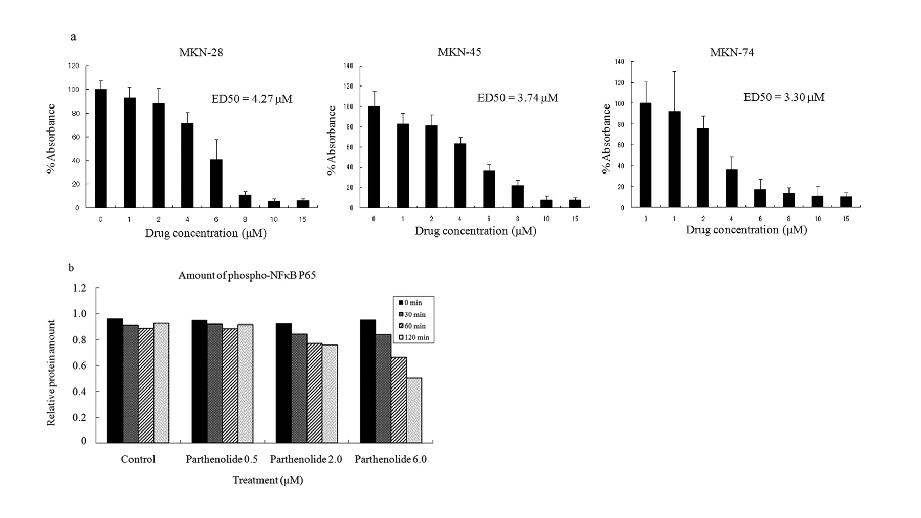
Effect of parthenolide on cell growth in vitro. The MTT assay was performed to examine the direct effect of parthenolide on cell growth using three gastric cancer cell lines, MKN-28, MKN-45, MKN-74. Parthenolide inhibited gastric cancer cell growth in a dose-dependent manner in all three gastric cell lines (Figure 1a). The efficacy of parthenolide in growth suppression was almost the same for these cell lines (ED 50=4.27, 3.74, and 3.30 μM in MKN-28, MKN-45, and MKN-74, respectively).

a: Growth inhibition with parthenolide in gastric cancer cell lines (MKN-28, MKN-45, and MKN-74) was examined with the MTT assay. Each cell line (5×10³ cells/well) was cultured for 96 hours with parthenolide at the concentration of, 0, 1, 2, 4, 6, 8, 10, and 15 μM. Data represent the mean±SEM of three different experiments performed in triplicate. b: Monitoring the activation of NF-κB by parthenolide on gastric cancer cell line (MKN-45). MKN-45 cells were treated with parthenolide at concentrations of 0, 0.5, 2.0, or 6.0 μM and the relative amount of NF-κB p65 phosphorylation was calculated at different time points.
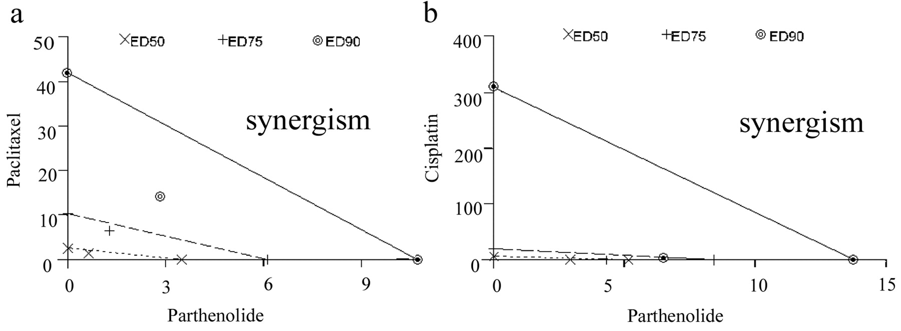
The analysis of isobolograms for the combination of parthenolide with antitumor agents: paclitaxel (a) and cisplatin (b) in three human gastric cancer cell lines (MKN-28, MKN-45, MKN-74). The synergic effect of parthenolide in combination with paclitaxel and cisplatin for MKN-45 cells is apparent.
Inhibition of phosphorylated NF-κB by parthenolide treatment. Parthenolide can suppress activation of NF-κB pathway in gastric cell line MKN-45 by monitoring NF-κB phosphorylation. MKN-45 cells were treated with parthenolide for 0, 30, 60, or 120 minutes at different concentrations (0, 0.5, 2.0, or 6.0 μM). According to the results, phospho-NF-κB p65 expression was suppressed in MKN-45 cells by parthenolide treatment in dose- and time-dependent manners (Figure 1b).
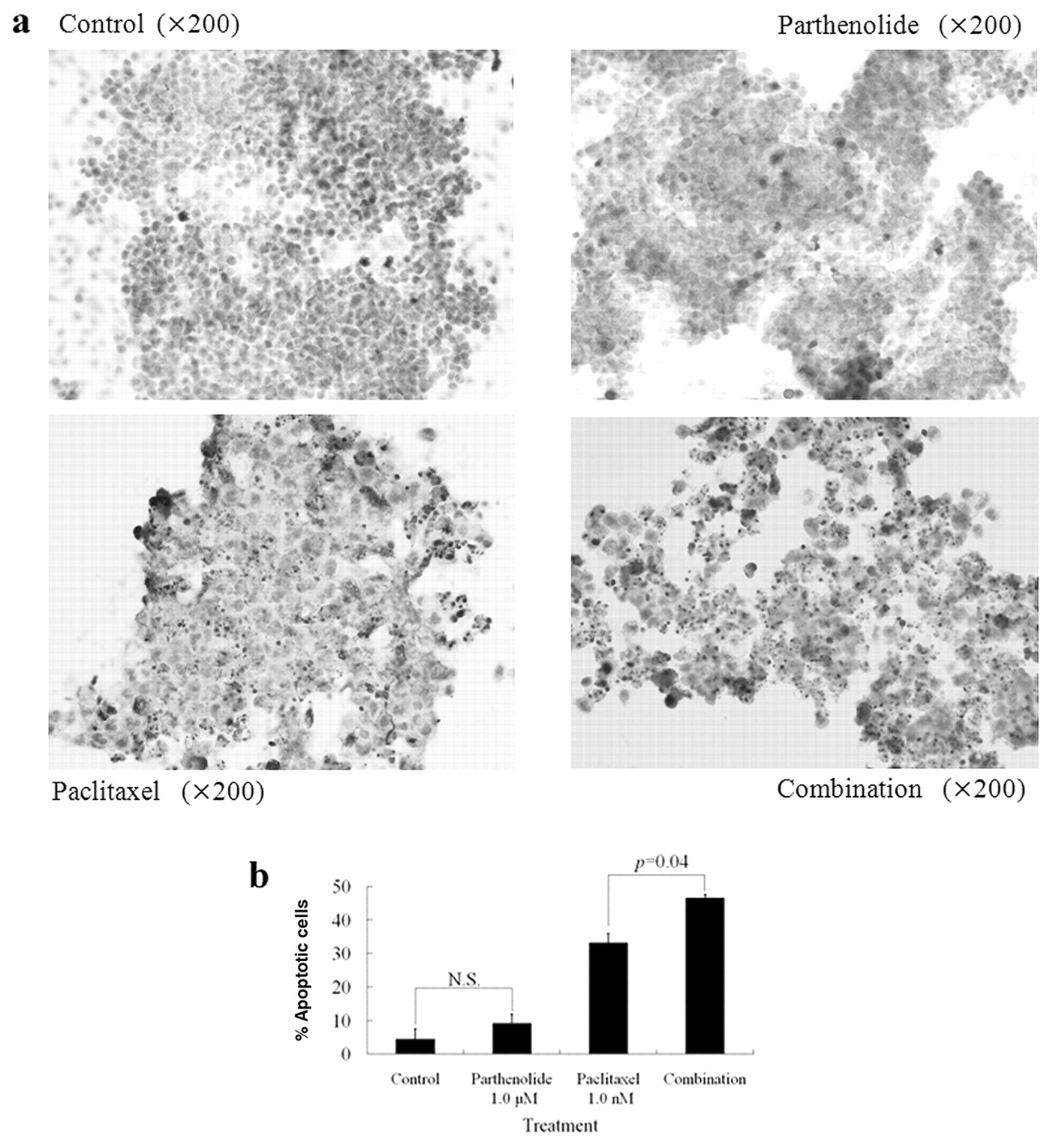
Apoptotic cells were evaluated by the percentage of TUNEL-positive cells in a ×400 field and the average of five fields was recorded as the apoptosis index. Data represent the mean±SEM. a: Immunohistochemical staining by the TUNEL method. b: Apoptosis index of each group treated with parthenolide and/or paclitaxel.
Identification of genes regulated by parthenolide treatment. We identified a panel of genes up- and down-regulated by treatment with parthenolide. Out of 84 genes associated with the NF-κB signaling pathway, 21 were down-regulated by >2-fold and 14 out of 21 were down-regulated by >3-fold (Table I). Most genes were down-regulated parthenolide treatment. There were only 10 genes up-regulated by an average fold-change of 1.27±0.22 and there were no genes up-regulated by >2-fold change (data not shown).
Increased sensitivity to antitumor agents with combination of parthenolide. We examined whether parthenolide could enhance the sensitivity of conventional antitumor agents. Paclitaxel and cisplatin were used as antitumor agents. Parthenolide tended to have a synergic antitumor effect in combination with paclitaxel and cisplatin in all three gastric cancer cell lines (Figure 2).
Effect of combination chemotherapy on apoptosis in cancer cells. According to the TUNEL experiments, the number of apoptotic cells increased in MKN-45 cells treated with paclitaxel compared to the control, however, there was no significant difference between parthenolide-treated and the control groups. On the other hand, the number of apoptotic cells was significantly higher in the combination group than in the paclitaxel-treated group (p=0.04) (Figure 3).

a: The inhibitory effect on peritoneal nodule formation by each treatment. Total surface area of all peritoneal nodules were scored and compared to that of the control (100%). b: The effect of parthenolide and or paclitaxel on survival duration in a mouse peritoneal dissemination model. Survival curves were drawn using the Kaplan-Meier method and comparisons of survival distribution were made using the log-rank test.
Effect of combination chemotherapy on peritoneal dissemination in the mouse model. The effect of combination chemotherapy was investigated in the peritoneal dissemination model. In this model, MKN-45-P cells were injected into the peritoneal cavity of each mouse. According to the previous animal experiment, we confirmed the formation of peritoneal dissemination on the day after intraperitoneal injection of MKN-45-P cells (29). The proportion of peritoneal nodules was significantly reduced in the group combination of parthenolide and paclitaxel treated with than in that treated with paclitaxel alone, in a dose dependent manner (Figure 4a).
Effect of combination chemotherapy on survival of mice with peritoneal dissemination. There was no significant difference between the survival curves of the control and the group treated with parthenolide alone at 4.0 mg/kg (p=0.5339). The group treated with paclitaxel alone at 1.0 mg/kg had a better prognosis than the control (p=0.0301). The combination of parthenolide at 0.25 mg/kg and paclitaxel conferred a better prognosis than for the control (p=0.0075), However, there was no significant improvement compared to the paclitaxel alone-treated group (p=0.4819). The combination of parthenolide at 4.0 mg/kg with paclitaxel gave a significantly better prognosis than for the group treated with paclitaxel alone (p=0.0405) (Figure 4b).
Toxicity of parthenolide and combination chemotherapy in the mouse model. To evaluate the toxic effect of parthenolide and combination chemotherapy, parthenolide only, combination of parthenolide and paclitaxel, or PBS were injected seven times into the peritoneal cavity. Such treatments produced no significant differences in body weight and food intake between these three groups. Furthermore, no abnormal pathology of major organs such as lung, heart, liver, spleen, and kidney was found (data not shown).
Discussion
Recent introduction of new active chemotherapeutic agents such as S-1 (a derivative of 5-FU), taxanes, platinum analogs, and CPT-11 have contributed to the improvement of patient survival in unresectable advanced gastric cancer in the world (31, 32). However, the therapeutic effect of chemotherapy is still limited due to the occurrence of resistance. Therefore, it may be a promising strategy to reduce chemoresistance to antitumor agents in solid tumors including gastric cancer. Chemotherapy is recognized to induce apoptosis as a therapeutic effect and chemoresistance likely involves an anti-apoptotic mechanism. NF-κB is a transcriptional activator for stimulation of cell growth and inhibition of apoptosis in cancer cell lines (33, 34). Uetsuka et al. showed that introduction of NF-κB decoy, which included κB-binding sites for p65 and p50, inhibited NF-κB activity and improved the chemosensitivity to 5-FU in a gastric cancer cell line (28). Parthenolide is recognized as one of the main sesquiterpene lactones responsible for the bioactivities of feverfew, which is commonly used as a food supplement for the treatment of migraines. Furthermore, feverfew reportedly has no severe side-effects (16) and a phase I trial of feverfew has been conducted for patients with cancer (37). Therefore, clinical application of feverfew should be safer and promising for patients with malignancies if the synergic effect with chemotherapy is confirmed.
Identification of genes down-regulated by parthenolide treatment.
In this study, we firstly investigated the effect of parthenolide on the NF-κB signaling pathway using gastric cancer cells. Firstly, we showed parthenolide itself significantly inhibited cell growth in three gastric cancer cell lines (MKN-28, MKN-45, and MKN-74). Furthermore, we confirmed that the phosphorylation of NF-κB was down-regulated by the treatment of parthenolide as shown by the ELISA assay. The synergic effect of parthenolide was confirmed in combination with paclitaxel and cisplatin in all three gastric cell lines. The combination treatment of paclitaxel and sublethal dose of parthenolide significantly induced apoptosis in MKN-45 gastric cancer cells compared to the group treated with paclitaxel alone. In the animal model, parthenolide significantly suppressed the formation of disseminated nodules of MKN-45-P gastric cancer cells as a single agent and also significantly enhanced the chemosensitivity to paclitaxel. Furthermore, the combined therapy of paclitaxel and higher dose (4.0 mg/kg) parthenolide significantly prolonged the survival duration. According to our results, parthenolide induced apoptosis and inhibited gastric cancer cell growth by suppression of NF-κB phosphorylation. Furthermore, parthenolide appears to be an effective and potent therapeutic agent for gastric cancer, both alone and in combination with chemotherapy.
This study also investigated the molecular mechanism of parthenolide using the PCR Array system which clarified the real-time PCR-based pathway-focused gene expression of 84 key genes related to NF-κB-mediated signal transduction. The experiment identified 21 down-regulated genes with >2-fold change and no genes were up-regulated by >2-fold by the treatment of parthenolide. Interestingly, most NF-κB-related genes were down-regulated. The most down-regulated gene was EST domain-containing protein Elk-1 (ELK1), a member of the ETS oncogene family (35). ELK-1 is the most prominent member of the ternary complex factor family of transcription factors. ELK1 activation occurs via a Ras/mitogen-activated protein kinase pathway (36). Ying et al. described that antisense oligonucleotide to ELK1 suppressed the tumorigenicity of human hepatocellular carcinoma cells (38). Our results indicated that suppression of tumor growth by parthenolide was partly due to the down-regulation of ELK1 and mitogen-activated protein kinase kinase kinase 1, which was also down-regulated by parthenolide in our study.
In conclusion, this study showed that parthenolide, recognized as one of the main components responsible for the bioactivities of feverfew, could be a candidate therapeutic agent, especially in combination with chemotherapy for the treatment of advanced gastric cancer.
Acknowledgments
We thank Dr Yutaka Yonemura for providing the MKN-45-P cell line.
- Received December 7, 2010.
- Revision received December 27, 2010.
- Accepted December 28, 2010.
- Copyright© 2011 International Institute of Anticancer Research (Dr. John G. Delinassios), All rights reserved


